. 2010 Jan;76(2):596-608.
doi: 10.1128/AEM.02037-09. Epub 2009 Nov 30.

Contributions of Francisella tularensis subsp. novicida chitinases and Sec secretion system to biofilm formation on chitin

Affiliations

Contributions of Francisella tularensis subsp. novicida chitinases and Sec secretion system to biofilm formation on chitin

Jeffrey J Margolis et al. Appl Environ Microbiol. 2010 Jan.

Abstract

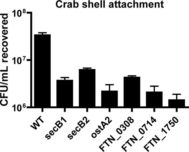
Francisella tularensis, the zoonotic cause of tularemia, can infect numerous mammals and other eukaryotes. Although studying F. tularensis pathogenesis is essential to comprehending disease, mammalian infection is just one step in the ecology of Francisella species. F. tularensis has been isolated from aquatic environments and arthropod vectors, environments in which chitin could serve as a potential carbon source and as a surface for attachment and growth. We show that F. tularensis subsp. novicida forms biofilms during the colonization of chitin surfaces. The ability of F. tularensis to persist using chitin as a sole carbon source is dependent on chitinases, since mutants lacking chiA or chiB are attenuated for chitin colonization and biofilm formation in the absence of exogenous sugar. A genetic screen for biofilm mutants identified the Sec translocon export pathway and 14 secreted proteins. We show that these genes are important for initial attachment during biofilm formation. We generated defined deletion mutants by targeting two chaperone genes (secB1 and secB2) involved in Sec-dependent secretion and four genes that encode putative secreted proteins. All of the mutants were deficient in attachment to polystyrene and chitin surfaces and for biofilm formation compared to wild-type F. novicida. In contrast, mutations in the Sec translocon and secreted factors did not affect virulence. Our data suggest that biofilm formation by F. tularensis promotes persistence on chitin surfaces. Further study of the interaction of F. tularensis with the chitin microenvironment may provide insight into the environmental survival and transmission mechanisms of this pathogen.


Figures

FIG. 1.
FIG. 1.
F. novicida biofilm formation on chitin surfaces. Images display SEM visualization of F. novicida colonization of crab shell pieces (A to D) and synthetic chitin films (E to H). Individual attached bacteria and small attached microcolonies were observed on crab shell pieces at 1 h (A and B). After 1 week, typical 3D biofilm architecture was observed, consisting of bacteria surrounded by an EPS matrix (C and D). Similar results were obtained after 1 h (E and F) and 1 week (G and H) on synthetic chitin.
FIG. 2.
FIG. 2.
Chitinase mutants are attenuated for chitin colonization in the absence of exogenous sugar. Stationary-phase wild-type (WT) and chitinase mutant bacteria were allowed to adhere for 1 h to crab shell pieces. Equivalently adherent strains were allowed to colonize these chitin surfaces in CDM with or without GlcNAc at 30°C. Triplicate samples were harvested 2 days postinoculation, and CFU were enumerated. F. novicida chitinase mutants (white) were recovered at statistically significantly lower levels than wild-type bacteria (black) (P < 0.001) when incubated in CDM (A) but in equivalent numbers in CDM with GlcNAc (B). Addition of the wild-type chiA and chiB genes to deletion mutant strains (grey; chiAcomp and chiBcomp) complemented the chitin colonization defects observed during colonization in CDM without GlcNAc (A).
FIG. 3.
FIG. 3.
Chitinase genes are required for biofilm architecture on chitin surfaces during nutrient stress. Images show representative colonization by wild-type and chitinase mutant strains on crab shells (A to D) or synthetic chitin films (E to H). Bacteria were allowed to attach for 1 h and then incubated for 1 week at 30°C before being processed for SEM. In contrast to the extensive 3D biofilm development in wild-type F. novicida, the chitinase mutants were present as single bacteria or small clusters of bacteria on both natural and synthetic chitin.
FIG. 4.
FIG. 4.
Francisella forms a mat-like biofilm under flow conditions. GFP-expressing F. novicida grown at room temperature (20 to 22°C) was imaged daily in flow cells run at 0.1 ml/min using CLSM. Representative images from triplicate experiments are shown. At 24 h (A), small groups of bacteria were present. Over the next 48 h (B and C), a uniform monolayer of bacteria was observed on the surface. By 96 h (D), depth in the biofilm was observed, and at 120 h (E), the biofilm reached an average thickness of 15 μm.
FIG. 5.
FIG. 5.
Kinetics of F. tularensis biofilm formation under static conditions. A modified O'Toole and Kolter assay was performed to compare the kinetics and relative levels of biofilm formation of F. novicida (solid circles) and an LVS (open circles). Bacterial growth (A and B) and crystal violet staining (C and D) were determined over time at 26°C (A and C) and 37°C (B and D) by OD570 readings. Both F. tularensis strains were found to acquire crystal violet stain at both temperatures. Growth and crystal violet staining were faster at 37°C for both strains.
FIG. 6.
FIG. 6.
Biofilm formation by virulent F. tularensis subsp. tularensis strains. F. novicida, an LVS, and type A strains SchuS4 and FT-10 were assayed for growth and crystal violet staining at 24 h postinoculation. Culture OD600 (A) and crystal violet staining (B) were determined after static growth at 37°C. F. novicida demonstrated increased growth kinetics and crystal violet staining compared to that of the other strains (P > 0.001). Virulent strains SchuS4 and FT-10 exhibited significantly higher crystal violet staining than the LVS.
FIG. 7.
FIG. 7.
Sec-secreted factors mediate initial attachment during biofilm formation. Five transposon insertions representing mutants defective in four genes in the Sec translocon (gray) and 18 transposon insertions representing mutants defective in 14 genes in putative secreted factors (white) identified in the forward genetic screen were tested in triplicate compared to wild-type (WT) F. novicida (black) for 8 h of biomass accumulation (A) and 1 h of initial attachment (B). Multiple transposon mutants were tested for genes identified more than once in the screen. Adherence of biomass at 8 h was used to measure biofilm formation. Attachment was assessed by crystal violet staining 1 h postinoculation of stationary-phase cultures. Targeted mutants defective in selected representative genes (white) showed similar defects in biofilm formation (C and E) and attachment (D and F) compared to wild-type F. novicida (black) when grown in MMH and CDM, respectively, based on crystal violet staining. Complementation of the deleted genes (gray) restored mutants to wild-type levels in all cases. Bars represent the means, and the lines indicate standard deviations calculated from triplicate samples of a representative experiment. Each experiment was repeated in triplicate. No data (ND) were obtained for FTN_0714 complementation due to technical difficulties.
FIG. 8.
FIG. 8.
The Sec translocon and secreted factors do not influence F. novicida virulence. Sec secretion-targeted F. tularensis deletion mutants were assessed for virulence in in vitro and in vivo models. Entry efficiency of F. novicida strains into RAW264.7 macrophage-like cells was measured as the percentage of the inoculum recovered from inside the cells at 30 min postinfection (A). Intracellular replication of wild-type (WT) and mutant bacteria was assessed as fold replication compared to 30-min counts at 8 h and 24 h postinfection (B). The ability of mutants to colonize the skin after i.d. inoculation (C) and the spleen after i.p. inoculation (D) was determined by determining CIs in C57BL/6J mice at 2 days postinfection. For all of the virulence assays, no difference was observed between the Sec secretion biofilm mutants and wild-type F. novicida.
FIG. 9.
FIG. 9.
Biofilm mutants are attenuated for attachment to chitin-based crab shell pieces. Stationary-phase cultures of secB1, secB2, ostA2, FTN_0308, FTN_0714, and FTN_1750 deletion mutants were allowed to attach for 1 h to sterile crab shell pieces. CFU of attached bacteria were enumerated in triplicate samples. Sec secretion biofilm mutants were found to attach statistically significantly less than wild-type (WT) F. novicida by unpaired t test (P > 0.01).

Similar articles

Cited by

References

    1. Adams, J. L., and R. J. C. McLean. 1999. Impact of rpoS deletion on Escherichia coli biofilms. Appl. Environ. Microbiol. 65:4285-4287. - PMC - PubMed
    1. Anda, P., J. Segura del Pozo, J. M. Díaz García, R. Escudero, F. J. García Peña, M. C. López Velasco, R. E. Sellek, M. R. Jiménez Chillarón, L. P. Sánchez Serrano, and J. F. Martínez Navarro. 2001. Waterborne outbreak of tularemia associated with crayfish fishing. Emerg. Infect. Dis. 7:575-582. - PMC - PubMed
    1. Balestrino, D., J. M. Ghigo, N. Charbonnel, J. A. Haagensen, and C. Forestier. 2008. The characterization of functions involved in the establishment and maturation of Klebsiella pneumoniae in vitro biofilm reveals dual roles for surface exopolysaccharides. Environ. Microbiol. 10:685-701. - PubMed
    1. Bartnicki-Garcia, S. 1968. Cell wall chemistry, morphogenesis, and taxonomy of fungi. Annu. Rev. Microbiol. 22:87-108. - PubMed
    1. Bassler, B. L., C. Yu, Y. C. Lee, and S. Roseman. 1991. Chitin utilization by marine bacteria. Degradation and catabolism of chitin oligosaccharides by Vibrio furnissii. J. Biol. Chem. 266:24276-24286. - PubMed

Publication types

LinkOut - more resources