. 2010 Feb;30(3):764-80.
doi: 10.1128/MCB.01458-09. Epub 2009 Nov 30.

Gene deletion mutants reveal a role for semaphorin receptors of the plexin-B family in mechanisms underlying corticogenesis

Affiliations

Gene deletion mutants reveal a role for semaphorin receptors of the plexin-B family in mechanisms underlying corticogenesis

A Hirschberg et al. Mol Cell Biol. 2010 Feb.

Abstract

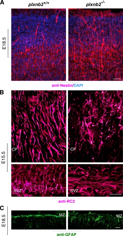
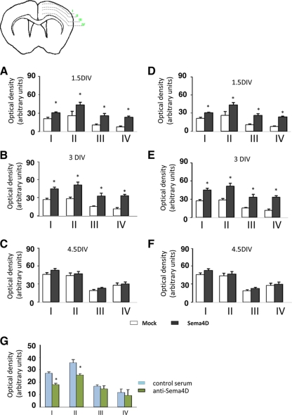
Semaphorins and their receptors, plexins, are emerging as key regulators of various aspects of neural and nonneural development. Semaphorin 4D (Sema4D) and B-type plexins demonstrate distinct expression patterns over critical time windows during the development of the murine neocortex. Here, analysis of mice genetically lacking plexin-B1 or plexin-B2 revealed the significance of Sema4D-plexin-B signaling in cortical development. Deficiency of plexin-B2 resulted in abnormal cortical layering and defective migration and differentiation of several subtypes of cortical neurons, including Cajal-Retzius cells, GABAergic interneurons, and principal cells in vivo. In contrast, a lack of plexin-B1 did not impact on cortical development in vivo. In various ex vivo assays on embryonic forebrain, Sema4D enhanced the radial and tangential migration of developing neurons in a plexin-B2-dependent manner. These results suggest that Sema4D-plexin-B2 interactions regulate mechanisms underlying cell specification, differentiation, and migration during corticogenesis.


Figures

FIG. 1.
FIG. 1.
Reciprocal expression of plexin-B1/B2 and Sema4d mRNAs, assessed via in situ hybridization during early stages of murine cortical development. Boxed areas are magnified in the inset. At E13.5, plexin-B1 mRNA (A) and plexin-B2 mRNA (B) are strongly expressed in the neuroepithelium (VZ and SVZ) whereas Sema4d is primarily found in the developing CP (C). Magnified views of expression in the developing neocortex are shown in panel D. At E13.5, the plexin-B1 and plexin-B2 mRNAs are strongly expressed in the neuroepithelium of the ganglionic eminence (GE) (E, F). Plexin-B2 and Sema4c mRNAs are expressed in the choroid plexus (G, H). Scale bars, 50 μm in all panels. Abbreviations: Ch.Pl, choroid plexus; CS, corpus striatum; NC, neocortex; LV, lateral ventricle; PP, preplate.
FIG. 2.
FIG. 2.
The architecture of the developing neocortex in mice lacking plexin-B1 (plxnb1−/−) is similar to that in their wild-type littermates (plxnb1+/+). Shown are representative images, derived on postnatal day 5, of immunoreactivity for various marker proteins in diverse populations of neurons and other cell types in the developing cortex, as well as BrdU birth-dating experiments. The nuclear stain DAPI (A, A′), the panneuronal marker NeuN (B, B′), cortical interneurons labeled via paralbumin (PV) (C, C′), Cajal-Retzius cells identified via antireelin immunohistochemistry analysis (D, D′), calretinin (CR) (E, E′), and development of astrocytes labeled via anti-GFAP immunoreactivity (F, F′) are similar in plexin-B1 deletion mutants (panels B′ to F′) and their wild-type littermates (B to F). (G, G′) Incorporation of BrdU in organotypic slice cultures of the developing cortex at E14.5 is comparable across plexin-B1 deletion mutants and wild-type embryos. LV, lateral ventricle. Scale bars, 50 μm in all panels.
FIG. 3.
FIG. 3.
Mice lacking plexin-B2 demonstrate a marked reduction in the density of neuronal populations in the developing CP at E18. (A) Decreased incidence of Tuj1-positive neurons in the CP of mice lacking plexin-B2 (plxnb2−/−) in comparison with their wild-type littermates (plxnb2+/+) at E18.5. Lower panels show staining of corresponding sections with the nuclear stain DAPI. (B and C) Typical examples (B) and quantification (C) of GABA-positive and GAD67-positive cells showing reduced numbers of GABAergic interneurons in the CP of mice lacking plexin-B2 at E18.5 in comparison with their wild-type littermates. Boxed areas are provided at a higher magnification in lower panels. *, P < 0.05 (Student's t test). Four embryos per genotype were tested. Scale bars, 25 μm.
FIG. 4.
FIG. 4.
Mice lacking plexin-B2 show reduced numbers of mature neurons in the developing CP at E18. (A, B) Abnormal distribution and reduced density of mature neurons identified via immunoreactivity to MAP2 and NeuN in the CP of mice lacking plexin-B2 in comparison with wild-type littermates at E18. Four embryos per genotype were tested.
FIG. 5.
FIG. 5.
Abnormal patterning and density of radial glia and progenitor cells, respectively, in the developing CP of mice lacking plexin-B2 (plxnb2−/−) in comparison with their wild-type littermate embryos (plxnb2+/+) at E18.5. (A) Nestin-positive neuronal and glial progenitors are found at a higher density in the developing CP of mice lacking plexin-B2 than in that of wild-type embryos at E18.5. (B) Radial glia, identified via RC2 immunoreactivity in the CP, are misformed and misaligned in mice lacking plexin-B2 at E15.5. Patterning of radial glia in the SVZ is shown in the lower panel. (C) Glial end feet identified via GFAP immunoreactivity at the basal laminae in the MZ are sporadically disrupted in mice lacking plexin-B2 at E18.5. Scale bars, 25 μm.
FIG. 6.
FIG. 6.
Analysis of Cajal-Retzius cells and the MZ in mice lacking plexin-B2 (plxnb2−/−) in comparison with their wild-type littermate embryos (plxnb2+/+) at E18.5. (A to D) Cajal-Retzius cells in the MZ, identified via reelin immunohistochemistry (A) or via expression of calretinin (C), are markedly reduced in the MZ of mice lacking plexin-B2 in comparison with their wild-type littermates. A quantitative summary is provided in panels B and D. (E) Nissl staining reveals that the packing density of cells in the MZ is markedly reduced in plxnb2−/− mutant mice compared with that of wild-type embryos. A quantitative summary is provided in panel F. *, P < 0.05 (Student's t test). Four embryos per genotype were tested. Scale bars, 25 μm.
FIG. 7.
FIG. 7.
Proliferation defects in the developing CP of mice lacking plexin-B2 (plxnb2−/−) in comparison to their wild-type littermate embryos (plxnb2+/+) at E18. (A, B) Nissl staining shows a marked hypoplasia of the CP, as well as the VZ, in plexin-B2 deletion mutants. A quantitative summary of the cell numbers/mm2 analyzed over the boxed areas is shown in panel B. (C) Mitogenic index of the germinal cells in the VZ, as identified via immunoreactivity for Ki-67 or PCNA, is reduced in plexin-B2 deletion mutants in comparison to that of their wild-type littermates. *, P < 0.05 (Student's t test). (D) Reduced density of anti-NeuN-immunoreactive neurons and (E) Nissl-stained cells in the SP of mice lacking plexin-B2. (F) Occurrence of anti-TAG1-immunoreactive afferents is reduced in the SP of plexin-B2 mutant mice compared to that in wild-type embryos. Scale bars, 75 μm and 50 μm in panels A to D, E, and F, respectively. LV, lateral ventricle.
FIG. 8.
FIG. 8.
Soluble Sema4D modulates migration, but not proliferation, of cells in VZ/SVZ explants and organotypic brain slice cultures derived from mice at E14.5. (A, B) Immunofluorescence images (A) and areas of migration (B) of Tuj1-positive cells out of VZ/SVZ explants cultured in the presence of either mock medium or Sema4D medium (130 explants per group). *, P < 0.05 (Student's t test). (C) Quantitative estimation of BrdU uptake in dissociated VZ/SVZ cells treated with mock medium, Sema4D medium, or EGF (50 nM). (D) Quantitative estimation of the total number of BrdU-labeled cells in the telencephalon in organotypic slices cultured in the presence of mock medium or Sema4D medium at 1.5, 3, or 4.5 DIV. (E) Typical examples of organotypic slice cultures treated with mock medium or Sema4D showing migration of BrdU-labeled precursors at various depths in the CP in the areas designated I to IV. (F) Quantitative analysis of numbers of BrdU-labeled cells in the areas designated I to IV in slices treated with mock medium or Sema4D, corresponding to the examples shown in panel E. Sema4D-treated cultures show an enhanced incidence of BrdU-labeled cells in the upper cortical layers, in contrast to the VZ (region I) (at least five or six slice cultures per treatment per DIV). *, P < 0.05 (ANOVA followed by Fisher's post hoc test). Scale bars, 30 μm.
FIG. 9.
FIG. 9.
Analysis of migration of newly born GABAergic neurons in organotypic slice cultures from mouse brain at E14.5 in the presence of mock medium or medium containing soluble Sema4D. (A) Schematic representation of an organotypic slice culture showing the regions designated I to IV in which GABA expression was studied. (B) Typical examples of immunofluorescence for GABA in the regions designated I, II, and III at 1.5, 3, and 4.5 DIV in the presence of mock medium or Sema4D. (C) Quantitative analysis of optical densities of GABA immunofluorescence in arbitrary units in the areas designated I to IV in the presence of mock medium or Sema4D (eight slice cultures per treatment per DIV). *, P < 0.05 (ANOVA followed by Fisher's post-hoc test). Scale bar in panel B, 25 μm.
FIG. 10.
FIG. 10.
Analysis of directionality of Sema4D effects on the migration of newly born GABAergic neurons in organotypic slice cultures from mouse brain at E14.5. The schematic represents an organotypic slice culture showing the regions designated I to IV in which GABA expression was studied. (A) HEK293 cell aggregates expressing soluble Sema4D were cocultured at the ventral aspect of organotypic slices, and the bar graphs represent optical densities of GABA immunofluorescence in the areas designated I to IV in slices cultured in the presence of mock medium or Sema4D. *, P < 0.05 (ANOVA followed by Fisher's post hoc test; five slices per group). (B) Sema4D released by HEK293 cell aggregates increases the migratory capacity of newly born neurons out of cocultured VZ/SVZ explants not only in the quadrant adjacent to the HEK aggregate (arrow) but also in other quadrants of the VZ/SVZ explants (arrowheads), thereby showing a lack of directional guidance. The panel on the right represents the mean area of migration out of the VZ explant. *, P < 0.05 (Student's t test; 30 explants per group).
FIG. 11.
FIG. 11.
Exogenously added, as well as endogenous, Sema4D promotes migration of newly born Tuj1-positive neurons in organotypic slice cultures from mouse brain at E14.5. The schematic represents an organotypic slice culture showing the regions designated I to IV in which Tuj1 immunoreactivity was studied. (A and B) Exogenous soluble Sema4D was either added to the culture matrix or released by HEK293 cell aggregates, which were cocultured at the ventral aspect of organotypic slices. The bar graphs represent the optical densities of Tuj1 immunofluorescence in the areas designated I to IV in slices cultured in the presence of mock medium or Sema4D (eight slice cultures per treatment). (C) Quantitative analysis of optical densities of Tuj1 immunofluorescence in the areas designated I to IV in the presence of control IgG or an antibody neutralizing endogenous Sema4D (six slices per treatment). *, P < 0.05 (ANOVA followed by post hoc Fisher test).
FIG. 12.
FIG. 12.
Role of endogenous Sema4D and ROCK activation in the migration of newly born GABAergic neurons in organotypic slice cultures from mouse brain at E14.5. The schematic represents an organotypic slice culture showing the regions designated I to IV in which GABA expression was studied. (A) Quantitative analysis of optical densities of GABA immunofluorescence in the areas designated I to IV in the presence of control IgG or an antibody neutralizing endogenous Sema4D (six slices per treatment). (B) Effects of treatment with the ROCK inhibitor Y27632 (3 μM) on GABAergic migration in organotypic slices cultured in the presence of exogenously added mock medium or Sema4D medium (eight slice cultures per treatment). *, P < 0.05 (ANOVA followed by Fisher's post hoc test).
FIG. 13.
FIG. 13.
The differential contribution of plexin-B1 and plexin-B2 to Sema4D-induced effects on migration in assays of VZ/SVZ explants and organotypic slice cultures of mouse embryonic cortex at E14.5. (A) In VZ/SVZ explants, Sema4D increases the migration of β-III-tubulin (Tuj1)-stained newly born neurons in mice lacking plexin-B1 (plxnb1−/−), as well as wild-type littermate controls (plxnb1+/+) (50 explants per treatment). (B) Sema4D-induced increase in the density of GABA-positive cells in various parts of the cortex (regions I to III) is maintained in mice lacking plexin-B1 (four or five slices per treatment per DIV). (C) Sema4D-induced increase in the migratory distance of BrdU-labeled cortical precursors in the upper cortical walls (regions II and III) is maintained in mice lacking plexin-B1 (four or five slices per treatment per DIV). (D) Sema4D-induced increase in the migratory capacity of DAPI+ cells out of VZ explants is lost in mice lacking plexin-B2 (plxnb2−/−). In contrast, promigratory effects of BDNF and Sema3A are preserved in explants derived from plxnb2−/− mutant mice (about 50 to 60 explants per treatment per genotype) *, P < 0.05 in all cases (ANOVA followed by Fisher's post hoc test). Scale bar in panel D, 50 μm.

Similar articles

Cited by

References

    1. Allendoerfer, K. L., and C. J. Shatz. 1994. The subplate, a transient neocortical structure: its role in the development of connections between thalamus and cortex. Annu. Rev. Neurosci. 17:185-218. - PubMed
    1. Andrews, W. D., M. Barber, and J. G. Parnavelas. 2007. Slit-Robo interactions during cortical development. J. Anat. 211:188-198. - PMC - PubMed
    1. Anton, E. S., M. A. Marchionni, K. F. Lee, and P. Rakic. 1997. Role of GGF/neuregulin signaling in interactions between migrating neurons and radial glia in the developing cerebral cortex. Development 124:3501-3510. - PubMed
    1. Aurandt, J., H. G. Vikis, J. S. Gutkind, N. Ahn, and K. L. Guan. 2002. The semaphorin receptor plexin-B1 signals through a direct interaction with the Rho-specific nucleotide exchange factor, LARG. Proc. Natl. Acad. Sci. U. S. A. 99:12085-12090. - PMC - PubMed
    1. Basile, J. R., T. Afkhami, and J. S. Gutkind. 2005. Semaphorin 4D/plexin-B1 induces endothelial cell migration through the activation of PYK2, Src, and the phosphatidylinositol 3-kinase-Akt pathway. Mol. Cell. Biol. 25:6889-6898. - PMC - PubMed

Publication types

MeSH terms