. 2010 Jan 5;107(1):355-60.
doi: 10.1073/pnas.0906186106. Epub 2009 Nov 30.

Prestimulus functional connectivity determines pain perception in humans

Affiliations

Prestimulus functional connectivity determines pain perception in humans

Markus Ploner et al. Proc Natl Acad Sci U S A. .

Abstract

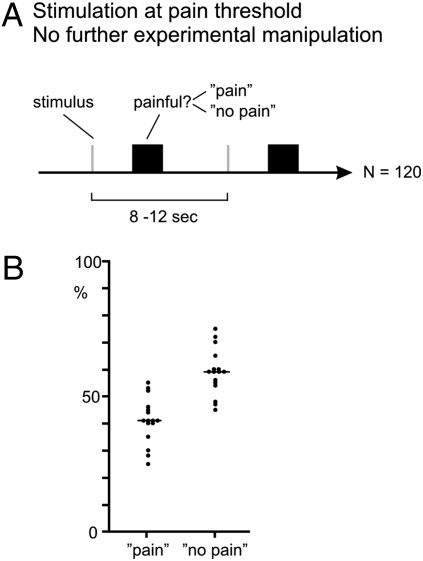
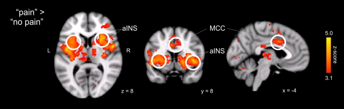
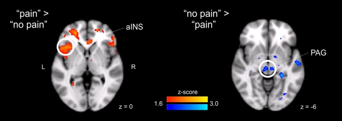
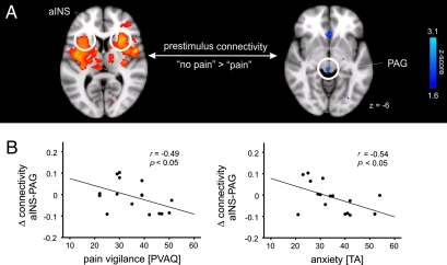
Pain is a highly subjective experience that can be substantially influenced by differences in individual susceptibility as well as personality. How susceptibility to pain and personality translate to brain activity is largely unknown. Here, we report that the functional connectivity of two key brain areas before a sensory event reflects the susceptibility to a subsequent noxious stimulus being perceived as painful. Specifically, the prestimulus connectivity among brain areas related to the subjective perception of the body and to the modulation of pain (anterior insular cortex and brainstem, respectively) determines whether a noxious event is perceived as painful. Further, these effects of prestimulus connectivity on pain perception covary with pain-relevant personality traits. More anxious and pain-attentive individuals display weaker descending connectivity to pain modulatory brain areas. We conclude that variations in functional connectivity underlie personality-related differences in individual susceptibility to pain.


Conflict of interest statement

The authors declare no conflict of interest.

Figures

Fig. 1.
Fig. 1.
Paradigm. (A) One hundred twenty brief radiant heat pulses were applied to the dorsal aspect of the right foot at an intensity around pain threshold without further experimental manipulation. After each trial, subjects indicated with a button press whether the stimulus was painful (pain) or not (no pain). Because stimulus intensity was kept constant throughout the experiment, differences in stimulus perception are likely to be at least partially attributable to fluctuations in the susceptibility to pain. (B) Individual and group mean proportion of pain and no pain trials.
Fig. 2.
Fig. 2.
Brain responses related to the subjective perception of pain. Brain areas in which neural responses are greater for pain than for no pain trials are shown. Activations are reported at a statistical threshold of P < 0.001, uncorrected, except for a priori hypothesized regions, which were thresholded at P < 0.005, uncorrected (see Materials and Methods). For visualization, activations are shown at a threshold of P < 0.001, uncorrected. aINS, anterior insular cortex; MCC, midcingulate cortex.
Fig. 3.
Fig. 3.
Differences in prestimulus neural activity before pain and no pain trials. (Left) Brain areas in which signals detected in a 3-sec prestimulus period were greater before pain trials than before no pain trials. (Right) Reverse contrast. Activations are reported at a statistical threshold of P < 0.001, uncorrected, except for a priori hypothesized regions, which were thresholded at P < 0.005, uncorrected (see Materials and Methods). For visualization, prestimulus activations are shown at a threshold of P < 0.05 uncorrected. aINS, anterior insular cortex (coordinates −40, 20, 2; z = 2.6); PAG (coordinates 4, −26, −8 and − 6, −24, −8; z = 2.6).
Fig. 4.
Fig. 4.
Prestimulus connectivity to the anterior insular cortex (aINS). (A) Differences in prestimulus connectivity to the bilateral aINS in a 3-sec prestimulus period as assessed by a PPI analysis. Brain areas whose connectivity to the anterior insular cortex is greater before no pain trials than before pain trials are shown. PAG (coordinates 4, −28, −4; z = 2.3). For coordinates of aINS, see Table 1. Voxels showing differences in connectivity to the left and right aINS were reported at a threshold of P < 0.05, small volume-corrected for the PAG (see Materials and Methods). For visualization, contrasts are shown at a threshold of P < 0.05 uncorrected. (B) Correlation between pain vigilance and anxiety and the difference between prestimulus insular and PAG connectivity. Pain vigilance and anxiety were assessed using the PVAQ, and the trait anxiety (TA) part of the STAI. Differences in connectivity between pain and no pain trials are given in effect sizes in arbitrary units. PAG coordinates were 0, −26, −16.

References

    1. Melzack R, Casey KL. Sensory, motivational, and central control determinants of pain: A new conceptual model. In: Kenshalo DRJ, editor. The Skin Senses. Springfield, IL: Charles C. Thomas; 1968. pp. 423–439.
    1. Boly M, et al. Baseline brain activity fluctuations predict somatosensory perception in humans. Proc Natl Acad Sci USA. 2007;104:12187–12192. - PMC - PubMed
    1. Deco G, Romo R. The role of fluctuations in perception. Trends Neurosci. 2008;31:591–598. - PubMed
    1. Crombez G, Van Damme S, Eccleston C. Hypervigilance to pain: An experimental and clinical analysis. Pain. 2005;116:4–7. - PubMed
    1. Rhudy JL, Meagher MW. The role of emotion in pain modulation. Curr Opin Psychiatry. 2001;14:241–245.

Publication types