

Cerebrovascular disease related to COL4A1 mutations in HANAC syndrome
- PMID: 19949034
- PMCID: PMC2881859
- DOI: 10.1212/WNL.0b013e3181c3fd12
Cerebrovascular disease related to COL4A1 mutations in HANAC syndrome
Abstract
Background: COL4A1 mutations cause familial porencephaly, infantile hemiplegia, cerebral small vessel disease (CSVD), and hemorrhagic stroke. We recently described hereditary angiopathy with nephropathy, aneurysm, and muscle cramps (HANAC) syndrome in 3 families with closely localized COL4A1 mutations. The aim of this study was to describe the cerebrovascular phenotype of HANAC.
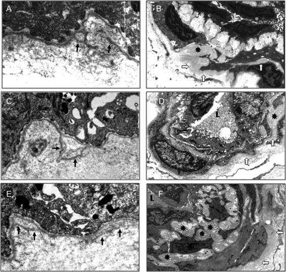
Methods: Detailed clinical data were collected in 14 affected subjects from the 3 families. MRI and magnetic resonance angiography (MRA) were performed in 9 of them. Skin biopsies were analyzed by electron microscopy in affected subjects in the 3 families.
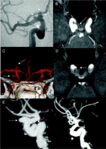
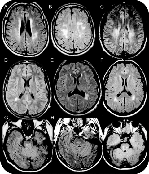
Results: Only 2 of 14 subjects had clinical cerebrovascular symptoms: a minor ischemic stroke at age 47 years and a small posttraumatic hemorrhage under anticoagulants at age 48 years. MRI-MRA showed cerebrovascular lesions in 8 of 9 studied subjects (mean age 39.4 years, 21-57 years), asymptomatic in 6 of them. Unique or multiple intracranial aneurysms, all on the carotid siphon, were observed in 5 patients. Seven patients had a CSVD characterized by white matter changes (7/7) affecting subcortical, periventricular, or pontine regions, dilated perivascular spaces (5/7), and lacunar infarcts (4/7). Infantile hemiplegia, major stroke, and porencephaly were not observed. Skin biopsies showed alterations of basement membranes at the dermoepidermal junction associated with expansion of extracellular matrix between smooth vascular cells in the arteriolar wall.
Conclusion: The cerebrovascular phenotype in hereditary angiopathy with nephropathy, aneurysm, and muscle cramps syndrome associates a cerebral small vessel disease and a large vessel disease with aneurysms of the carotid siphon. It is consistent with a lower susceptibility to hemorrhagic stroke than in familial porencephaly, suggesting an important clinical heterogeneity in the phenotypic expression of disorders related to COL4A1 mutations.
Figures
References
-
- Mayne R. Collagenous proteins of blood vessels. Arteriosclerosis 1986;6:585–593. - PubMed
-
- Shekhonin BV, Domogatsky SP, Muzykantov VR, et al. distribution of type I, III, IV and V collagen in normal and atherosclerotic human arterial wall: immunomorphological characteristics. Coll Relat Res 1985;5:355–368. - PubMed
-
- Urabe N, Naito I, Saito K, et al. Basement membrane type IV collagen molecules in the choroids plexus, pia mater and capillaries in the mouse brain. Arch Histol Cytol 2002;65:133–143. - PubMed
-
- Gould DB, Phalan FC, Breedveld GJ, et al. Mutations in Col4a1 cause perinatal cerebral hemorrhage and porencephaly. Science 2005;308:1167–1171. - PubMed
-
- Mancini GMS, De Coo IFM, Lequin MH, Arts WF. Hereditary porencephaly: clinical and MRI findings in two Dutch families. Eur J Paediatr Neurol 2004;8:45–54. - PubMed
Publication types
MeSH terms
Substances
Grants and funding
LinkOut - more resources
Full Text Sources
Medical
Molecular Biology Databases