. 2009 Nov 20:3:14.
doi: 10.3389/neuro.03.014.2009. eCollection 2009.

Implications of functional anatomy on information processing in the deep cerebellar nuclei

Affiliations

Implications of functional anatomy on information processing in the deep cerebellar nuclei

Yuval Baumel et al. Front Cell Neurosci. .

Abstract

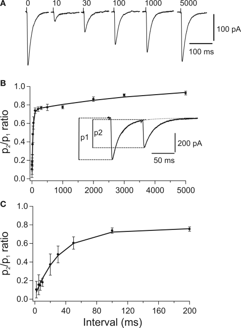
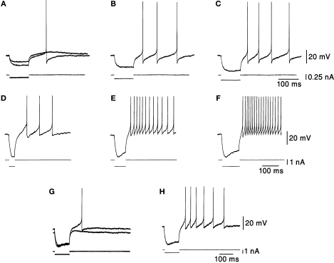
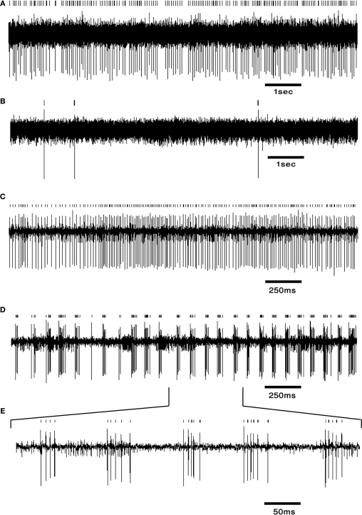
The cerebellum has been implicated as a major player in producing temporal acuity. Theories of cerebellar timing typically emphasize the role of the cerebellar cortex while overlooking the role of the deep cerebellar nuclei (DCN) that provide the sole output of the cerebellum. Here we review anatomical and electrophysiological studies to shed light on the DCN's ability to support temporal pattern generation in the cerebellum. Specifically, we examine data on the structure of the DCN, the biophysical properties of DCN neurons and properties of the afferent systems to evaluate their contribution to DCN firing patterns. In addition, we manipulate one of the afferent structures, the inferior olive (IO), using systemic harmaline injection to test for a network effect on activity of single DCN neurons in freely moving animals. Harmaline induces a rhythmic firing pattern of short bursts on a quiescent background at about 8 Hz. Other neurons become quiescent for long periods (seconds to minutes). The observed patterns indicate that the major effect harmaline exerts on the DCN is carried indirectly by the inhibitory Purkinje cells (PCs) activated by the IO, rather than by direct olivary excitation. Moreover, we suggest that the DCN response profile is determined primarily by the number of concurrently active PCs, their firing rate and the level of synchrony occurring in their transitions between continuous firing and quiescence. We argue that DCN neurons faithfully transfer temporal patterns resulting from strong correlations in PCs state transitions, while largely ignoring the timing of simple spikes from individual PCs. Future research should aim at quantifying the contribution of PC state transitions to DCN activity, and the interplay between the different afferent systems that drive DCN activity.

Keywords: cerebellar nuclei; chronic recording; harmaline; inferior olive; rebound firing; short-term depression; temporal patterns.


Figures

Figure 1
Figure 1
Paired pulse depression (PPD) of compound PC-DCN IPSCs. (A) Representative example of IPSCs evoked by paired stimulation. Each trace illustrates the average of 25 single IPSCs evoked in a DCN by pairs of stimuli with different intervals applied to PC axons. Number above each trace indicates the interpulse interval in milliseconds. Traces corresponding to 10 and 30 ms were obtained after subtracting the response to the 1st stimulus (labeled with “0”). (B,C) Time course of recovery from PPD shown at two different time scales. Average peak amplitude ratio (p2/p1) was plotted against the interpulse interval (n = 24). The curves represent a double exponential expression fitted to the data. The peak amplitude of IPSCs was measured from the baseline to the peak of the respective IPSCs. Taken with permission from Pedroarena and Schwarz (2003).
Figure 2
Figure 2
Responses to hyperpolarizing current pulses. (A–C) Rebound responses were seen after hyperpolarizing current pulses. The smallest pulse produced only a subthreshold depolarization. As the stimulus increased in amplitude an increasing number of action potentials were generated. (D–F) The responses were dependent on the duration of preceding hyperpolarization. More spikes were seen after long hyperpolarizations. (G,H) The rebound response was dependent on the initial membrane potential. The response was larger when the membrane was depolarized. Taken with permission from Jahnsen (1986).
Figure 3
Figure 3
DCN neurons exhibit typical firing patterns following harmaline injection. (A,B) An example of a tonically firing neuron (A) that became almost completely quiescent after harmaline injection (B). (C,D) An example of a tonically firing neuron (C) that exhibited an ON/OFF firing pattern after harmaline injection (D). (E) Time expansion of the marked section in (D) showing the typical cycle of the ON/OFF pattern. Small bars above the trace mark the sorted spikes. The surgical procedure has been described in detail (Cohen and Nicolelis, ; Jacobson et al., 2009). In brief, adult Long Evans male rats weighing 350–500 g (Harlan, Indianapolis, IN, USA) were initially sedated by 5% isoflurane and then injected i.m. with ketamine HCl and xylazine HCl (100 and 10 mg/kg, respectively). Supplementary injections of ketamine and xylazine were given as required. The skull surface was exposed and a craniotomy, slightly larger than the electrode array, was performed above the medial and interposed nuclei. Center of implant was at −11.3 mm posterior to bregma, 1.5 mm lateral to the midline (coordinates taken from Paxinos and Watson, 1998). Arrays of 16 electrodes were lowered 4 mm from the surface of the brain and fixed using dental cement. Rats were allowed at least 2 weeks of recovery prior to recording. Electrode location was verified using histology.

References

    1. Aizenman C. D., Huang E. J., Linden D. J. (2003). Morphological correlates of intrinsic electrical excitability in neurons of the deep cerebellar nuclei. J. Neurophysiol. 89, 1738–174710.1152/jn.01043.2002 - DOI - PubMed
    1. Aizenman C. D., Linden D. J. (1999). Regulation of the rebound depolarization and spontaneous firing patterns of deep nuclear neurons in slices of rat cerebellum. J. Neurophysiol. 82, 1697–1709 - PubMed
    1. Aksenov D., Serdyukova N., Irwin K., Bracha V. (2004). GABA neurotransmission in the cerebellar interposed nuclei: involvement in classically conditioned eyeblinks and neuronal activity. J. Neurophysiol. 91, 719–72710.1152/jn.00859.2003 - DOI - PubMed
    1. Aksenov D. P., Serdyukova N. A., Bloedel J. R., Bracha V. (2005). Glutamate neurotransmission in the cerebellar interposed nuclei: involvement in classically conditioned eyeblinks and neuronal activity. J. Neurophysiol. 93, 44–5210.1152/jn.00586.2004 - DOI - PubMed
    1. Alvina K., Walter J. T., Kohn A., Ellis-Davies G., Khodakhah K. (2008). Questioning the role of rebound firing in the cerebellum. Nat. Neurosci. 11, 1256–125810.1038/nn.2195 - DOI - PMC - PubMed