Dissociable effects of dopamine on neuronal firing rate and synchrony in the dorsal striatum
- PMID: 19949467
- PMCID: PMC2784296
- DOI: 10.3389/neuro.07.028.2009
Dissociable effects of dopamine on neuronal firing rate and synchrony in the dorsal striatum
Abstract
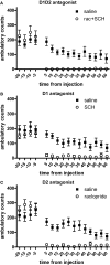
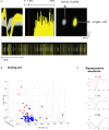
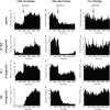
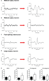
Previous studies showed that dopamine depletion leads to both changes in firing rate and in neuronal synchrony in the basal ganglia. Since dopamine D1 and D2 receptors are preferentially expressed in striatonigral and striatopallidal medium spiny neurons, respectively, we investigated the relative contribution of lack of D1 and/or D2-type receptor activation to the changes in striatal firing rate and synchrony observed after dopamine depletion. Similar to what was observed after dopamine depletion, co-administration of D1 and D2 antagonists to mice chronically implanted with multielectrode arrays in the striatum caused significant changes in firing rate, power of the local field potential (LFP) oscillations, and synchrony measured by the entrainment of neurons to striatal local field potentials. However, although blockade of either D1 or D2 type receptors produced similarly severe akinesia, the effects on neural activity differed. Blockade of D2 receptors affected the firing rate of medium spiny neurons and the power of the LFP oscillations substantially, but it did not affect synchrony to the same extent. In contrast, D1 blockade affected synchrony dramatically, but had less substantial effects on firing rate and LFP power. Furthermore, there was no consistent relation between neurons changing firing rate and changing LFP entrainment after dopamine blockade. Our results suggest that the changes in rate and entrainment to the LFP observed in medium spiny neurons after dopamine depletion are somewhat dissociable, and that lack of D1- or D2-type receptor activation can exert independent yet interactive pathological effects during the progression of Parkinson's disease.
Keywords: Parkinson's disease; caudate; entrainment; local field potentials; movement; oscillations; putamen.
Figures


Similar articles
-
Dopaminergic treatment weakens medium spiny neuron collateral inhibition in the parkinsonian striatum.J Neurophysiol. 2017 Mar 1;117(3):987-999. doi: 10.1152/jn.00683.2016. Epub 2016 Dec 7. J Neurophysiol. 2017. PMID: 27927785 Free PMC article.
-
Selective Vulnerability of Striatal D2 versus D1 Dopamine Receptor-Expressing Medium Spiny Neurons in HIV-1 Tat Transgenic Male Mice.J Neurosci. 2017 Jun 7;37(23):5758-5769. doi: 10.1523/JNEUROSCI.0622-17.2017. Epub 2017 May 4. J Neurosci. 2017. PMID: 28473642 Free PMC article.
-
L-DOPA Oppositely Regulates Synaptic Strength and Spine Morphology in D1 and D2 Striatal Projection Neurons in Dyskinesia.Cereb Cortex. 2016 Oct 17;26(11):4253-4264. doi: 10.1093/cercor/bhw263. Cereb Cortex. 2016. PMID: 27613437 Free PMC article.
-
D1 and D2 dopamine-receptor modulation of striatal glutamatergic signaling in striatal medium spiny neurons.Trends Neurosci. 2007 May;30(5):228-35. doi: 10.1016/j.tins.2007.03.008. Epub 2007 Apr 3. Trends Neurosci. 2007. PMID: 17408758 Review.
-
Segregation of D1 and D2 dopamine receptors in the striatal direct and indirect pathways: An historical perspective.Front Synaptic Neurosci. 2023 Jan 19;14:1002960. doi: 10.3389/fnsyn.2022.1002960. eCollection 2022. Front Synaptic Neurosci. 2023. PMID: 36741471 Free PMC article. Review.
Cited by
-
Longitudinal Changes in Isolated Rapid Eye Movement Sleep Behavior Disorder-Related Metabolic Pattern Expression.Mov Disord. 2021 Aug;36(8):1889-1898. doi: 10.1002/mds.28592. Epub 2021 Mar 31. Mov Disord. 2021. PMID: 33788284 Free PMC article.
-
Dopaminergic modulation of the striatal microcircuit: receptor-specific configuration of cell assemblies.J Neurosci. 2011 Oct 19;31(42):14972-83. doi: 10.1523/JNEUROSCI.3226-11.2011. J Neurosci. 2011. PMID: 22016530 Free PMC article.
-
Orbitofrontal and striatal circuits dynamically encode the shift between goal-directed and habitual actions.Nat Commun. 2013;4:2264. doi: 10.1038/ncomms3264. Nat Commun. 2013. PMID: 23921250 Free PMC article.
-
Effects of dopamine depletion on LFP oscillations in striatum are task- and learning-dependent and selectively reversed by L-DOPA.Proc Natl Acad Sci U S A. 2012 Oct 30;109(44):18126-31. doi: 10.1073/pnas.1216403109. Epub 2012 Oct 16. Proc Natl Acad Sci U S A. 2012. PMID: 23074253 Free PMC article.
-
An aetiological Foxp2 mutation causes aberrant striatal activity and alters plasticity during skill learning.Mol Psychiatry. 2012 Nov;17(11):1077-85. doi: 10.1038/mp.2011.105. Epub 2011 Aug 30. Mol Psychiatry. 2012. PMID: 21876543 Free PMC article.
References
-
- Bergman H., Wichmann T., Karmon B., DeLong M. R. (1994). The primate subthalamic nucleus. II. Neuronal activity in the MPTP model of Parkinsonism. J. Neurophysiol. 72, 507–520 - PubMed
LinkOut - more resources
Full Text Sources

