

Repair of extensive perineal hypospadias in a Boston terrier using tubularized incised plate urethroplasty
- PMID: 19949553
- PMCID: PMC2726019
Repair of extensive perineal hypospadias in a Boston terrier using tubularized incised plate urethroplasty
Abstract
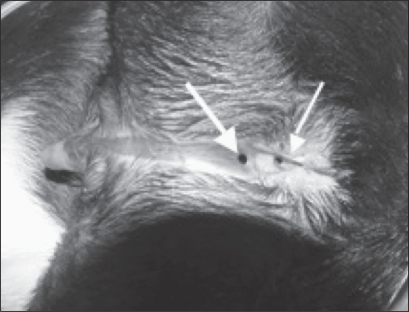
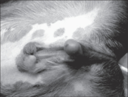
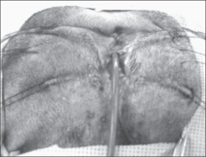
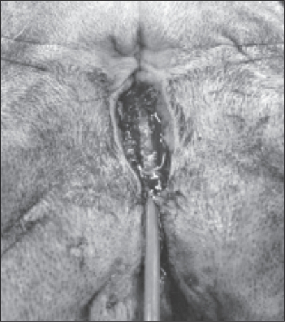
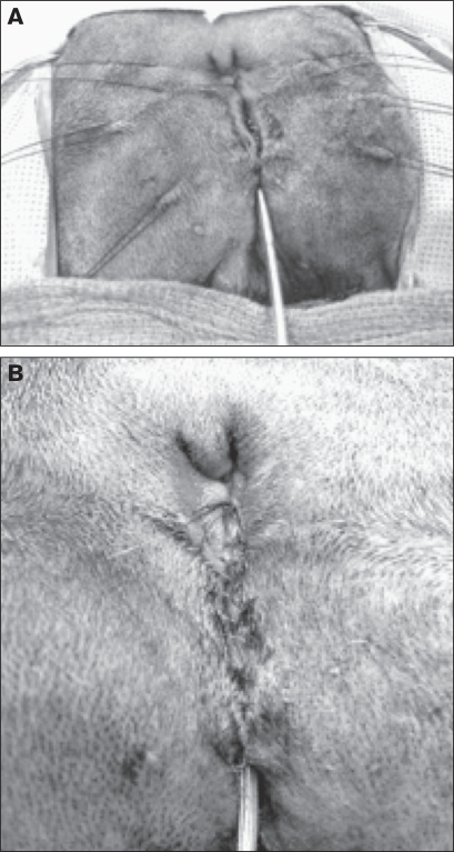
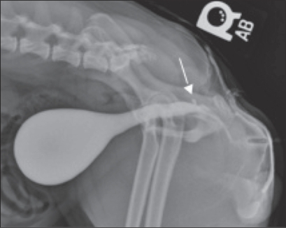
This report provides a detailed description of the surgical repair of perineal hypospadias in a Boston terrier. A 2-year patient follow-up, including diagnostic data demonstrating urethral patency and resolution of recurrent urocystitis and urine skin scald are reported.
Réparation d’un hypospadias périnéal important chez un Boston terrier en utilisant l’urétroplastie à lame gravée tubulaire. Ce rapport présente une description détaillée de la réparation chirurgicale d’un hypospadias périnéal chez un Boston terrier. Un rapport est présenté sur un suivi de 2 ans chez un patient qui inclut des données diagnostiques démontrant la perméabilité urétrale et la résolution des rechutes de cystites et de taches dermiques causées par l’urine.
(Traduit par Isabelle Vallières)
Figures

References
-
- Hobson HP. Penis and prepuce. In: Bojrab MJ, editor. Current Techniques in Small Animal Surgery. 4th ed. Philadelphia: Williams and Wilkins; 1998. p. 528.
-
- Boothe HW. Penis, prepuce, and scrotum. In: Slatter D, editor. Textbook of Small Animal Surgery. 3rd ed. Philadelphia: WB Saunders; 2003. pp. 1535–1536.pp. 1640–1641.
-
- Hayes HM, Wilson GP. Hospital incidence of hypospadias in dogs in North America. Vet Rec. 1986;118:605–606. - PubMed
-
- Hedlund CS. Surgery of the male reproductive tract. In: Fossum TW, editor. Small Animal Surgery. 2nd ed. St. Louis: Mosby; 2002. pp. 662–665.
-
- Croshaw JE, Jr, Brodey RS. Failure of preputial closure in a dog. J Am Vet Med Assoc. 1960;136:450–452. - PubMed
Publication types
MeSH terms
LinkOut - more resources
Full Text Sources