

doi: 10.4070/kcj.2009.39.5.209.
Epub 2009 May 28.
A case of aortopulmonary fistula caused by a huge thoracic aortic aneurysm
Affiliations
- PMID: 19949581
- PMCID: PMC2771785
- DOI: 10.4070/kcj.2009.39.5.209
Item in Clipboard
A case of aortopulmonary fistula caused by a huge thoracic aortic aneurysm
Korean Circ J.
2009 May.
Abstract
Aortopulmonary fistula is an uncommon but often fatal condition resulting as a late complication of an aortic aneurysm. The most common cause is erosion of a false aneurysm of the descending thoracic aorta into the pulmonary artery, resulting in the development of a left-to-right shunt and leading to acute pulmonary edema and right heart failure. We report an our experience with aortopulmonary fistula as a rare complication associated with thoracic aortic aneurysm and high output heart failure.
Keywords: Aortic aneurysm; Fistula; Heart failure.
Figures
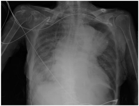
Chest AP showed cardiomegaly with right pleural effusion. An aneurysm of the arch of the aorta is seen, causing mediastinal widening. AP: anterioposterior.
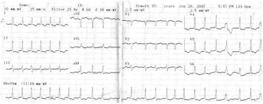
The electrocardiogram showed atrial fibrillation, left ventricular hypertrophy and T wave inversion accompanied by ST-segment depression in V3-V6.
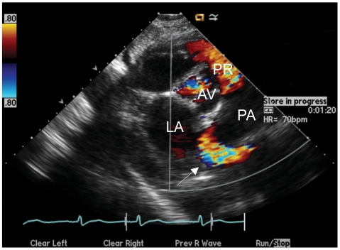
At the aortic root level on the parasternal short axis view (color doppler) showing abnormal blood flow around the pulmonary artery (white arrow). AV: aortic valve, PR: pulmonic regurgitation, LA: Left atrium, PA: pulmonary artery.
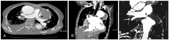
The CT scan shows thrombosed aortic aneurysm (white arrow) and aortic aneurysm is communicated with pulmonary artery resulting in aortopulmonary fistula (black arrow) (A). coronal reformated image demonstrates communication from aortic aneurysm to main pulmonary artery (black arrow) (B). 3D reconstructed image shows the aortopulmonary fistula (black arrow) (C).
References
-
- Bickerstaff LK, Pairolero PC, Hollier LH, et al. Thoracic aortic aneurysms: a population based study. Surgery. 1982;92:1103–1108. - PubMed
-
- Zips DP, Libby P, Bonow RO, Braunwald E. Braunwald's Heart disease: A textbook of cardiovascular medicine. 7th ed. Philadelphia: Sauders; 2005. pp. 1410–1415.
-
- Belgi A, Altekin E, Yalcinkaya S, Tüzüner FE. Acquired aortopulmonary fistula: a case of ruptured aneurysm of the thoracic aorta. Anadolu Kardiyol Derg. 2003;3:275–278. - PubMed
-
- Halperin JL, Olin JW. Disease of the aorta. In: Fuster V, editor. Hurst's The Heart. 11th ed. New York: McGraw-Hil; 2004. p. 2304.
LinkOut - more resources
Full Text Sources
