

How does the left ventricle work? Ventricular rotation as a new index of cardiac performance
- PMID: 19949617
- PMCID: PMC2771832
- DOI: 10.4070/kcj.2009.39.9.347
How does the left ventricle work? Ventricular rotation as a new index of cardiac performance
Abstract
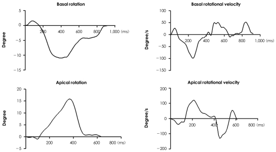
Although simple cylindrical or ellipsoidal left ventricular (LV) geometry with transverse or circumferential muscle contraction has been traditionally used to estimate LV performance, the estimated LV ejection fraction (EF) with muscle fiber shortening up to 20% is less than 50% of maximum, which is lower than the normal EF observed in routine clinical practice. Thus, oblique fiber orientation and LV rotation, in addition to radial thickening and longitudinal shortening, is predicted as an essential component of effective LV pumping. This was confirmed by animal experiments using surgically implanted markers or invasive sonomicrometry. Demonstration of the muscle band extending from the pulmonary artery to the aorta, which connects the ventricular myocardium, both right ventricle and LV as a continuous band (muscle band theory) provides an anatomical backbone of helical configuration of the cardiac muscle band with descending and ascending segments wrapping the LV apex. Moreover, sequential, non-simultaneous, activation and contraction of the helicoids muscle band contributes to LV rotation or twist motion. Recently, magnetic resonance imaging and speckle tracking echocardiography (STE) techniques have provided an excellent noninvasive way to measure LV rotation and twist, which is expected to contribute to a more thorough evaluation of both LV systolic and diastolic function. Initial animal experiments showed that quantification of apical rotation or LV twist using STE is more accurate for estimating LV systolic function than conventional EF under a variety of LV inotropic conditions, irrespective of coronary ligation. As de-rotation or the untwisting rate can also be measured by STE, the role of ventricular untwisting as a temporal link between LV relaxation and suction can be addressed. Further clinical investigations are needed to determine the real clinical impact of these new indices of LV mechanical function.
Keywords: Echocardiography; Rotation; Ventricular function.
Figures


References
-
- Ingels NB., Jr Myocardial fiber architecture and left ventricular function. Technol Health Care. 1997;5:45–52. - PubMed
-
- Lower R. Tractus de corde. In: Gunther RT, editor. Early Science in Oxford. Reprint. Oxford, UK: Dawsons, Pall Mall, London; 1968. p. 1669.
-
- Ingels NB, Daughters GT, 2nd, Stinson EB, Alderman EL. Measurement of midwall myocardial dynamics in intact man by radiography of surgically implanted markers. Circulation. 1975;52:859–867. - PubMed
-
- Torrent-Guasp F, Ballester M, Buckberg GD, et al. Spatial orientation of the ventricular muscle band: physiologic contribution and surgical implications. J Thorac Cardiovasc Surg. 2001;122:389–392. - PubMed
LinkOut - more resources
Full Text Sources
Research Materials
Miscellaneous
