

The best predictor for right ventricular dysfunction in acute pulmonary embolism: comparison between electrocardiography and biomarkers
- PMID: 19949622
- PMCID: PMC2771828
- DOI: 10.4070/kcj.2009.39.9.378
The best predictor for right ventricular dysfunction in acute pulmonary embolism: comparison between electrocardiography and biomarkers
Abstract
Background and objectives: Right ventricular (RV) dysfunction is associated with a poor prognosis in patients with an acute pulmonary embolism (APE). We studied the role of electrocardiography and biomarkers for early detection and recovery of right ventricular dysfunction (RVD) in APE.
Subjects and methods: The medical records of 48 consecutive patients diagnosed with APE using CT-angiography, at the Kangdong Sacred Heart Hospital, between January 2004 and February 2008 were reviewed retrospectively. RVD was assessed by serial echocardiography (ECG). Patients with one of the following were considered to have RVD: 1) RV dilatation (enddiastolic diameter >30 mm in the parasternal long axis view), 2) RV free wall hypokinesia, and 3) paradoxical septal systolic motion. We compared the electrocardiographic findings and the biomarkers for the early detection of RVD.
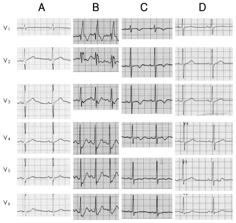
Results: The electrocardiographic findings showed T-wave inversion (TWI) in leads V1 to V3 with a sensitivity of 75% and a specificity of 95%, and a diagnostic accuracy of 80% for the detection of RVD, with positive and negative predictive values of 95.5% and 73.1%, respectively; these results were better than the biomarkers such as cardiac enzymes or B-type natriuretic peptide (BNP) for the early detection of RVD. TWIs persisted throughout the period of RVD, in contrast to a transient S1Q3T3 pattern detected during the acute phase only.
Conclusion: TWIs in leads V1 to V3 had the greatest sensitivity and diagnostic accuracy for early detection of RVD, and normalization of the TWIs was associated with recovery of RVD in APE.
Keywords: Electrocardiography; Pulmonary embolism; Ventricular dysfunction, right.
Figures
Similar articles
-
Normalization of negative T-wave on electrocardiography and right ventricular dysfunction in patients with an acute pulmonary embolism.Korean J Intern Med. 2012 Mar;27(1):53-9. doi: 10.3904/kjim.2012.27.1.53. Epub 2012 Feb 28. Korean J Intern Med. 2012. PMID: 22403500 Free PMC article.
-
A new electrocardiographic parameter terminal D1S + D3R predicts right ventricular dilatation in acute pulmonary embolism.Acta Cardiol. 2024 Nov;79(9):1021-1029. doi: 10.1080/00015385.2024.2396760. Epub 2024 Sep 17. Acta Cardiol. 2024. PMID: 39286922
-
Predictive value of reduced pulmonary arterial elasticity in acute pulmonary embolism for right ventricular dysfunction.J Thromb Thrombolysis. 2023 Nov;56(4):529-537. doi: 10.1007/s11239-023-02873-z. Epub 2023 Aug 7. J Thromb Thrombolysis. 2023. PMID: 37548900
-
ST-segment elevation in V1-V4 in acute pulmonary embolism: a case presentation and review of literature.Eur Heart J Acute Cardiovasc Care. 2016 Dec;5(8):579-586. doi: 10.1177/2048872615604273. Epub 2015 Sep 15. Eur Heart J Acute Cardiovasc Care. 2016. PMID: 26373811 Review.
-
B-type natriuretic peptide in acute pulmonary embolism.Clin Chim Acta. 2008 Dec;398(1-2):1-4. doi: 10.1016/j.cca.2008.07.020. Epub 2008 Jul 24. Clin Chim Acta. 2008. PMID: 18706401 Review.
Cited by
-
The Zurkurnai ECG Pattern: A Novel ECG Pattern of the High-Risk Features of Acute Pulmonary Embolism.Cureus. 2024 Jan 24;16(1):e52889. doi: 10.7759/cureus.52889. eCollection 2024 Jan. Cureus. 2024. PMID: 38274596 Free PMC article.
-
Correlation between ST-segment elevation and negative T waves in the precordial leads in acute pulmonary embolism: insights into serial electrocardiogram changes.Ann Noninvasive Electrocardiol. 2014 Jul;19(4):398-405. doi: 10.1111/anec.12115. Epub 2013 Nov 8. Ann Noninvasive Electrocardiol. 2014. PMID: 24206526 Free PMC article.
-
Normalization of negative T-wave on electrocardiography and right ventricular dysfunction in patients with an acute pulmonary embolism.Korean J Intern Med. 2012 Mar;27(1):53-9. doi: 10.3904/kjim.2012.27.1.53. Epub 2012 Feb 28. Korean J Intern Med. 2012. PMID: 22403500 Free PMC article.
-
Assessment and diagnosis of right ventricular failure-retrospection and future directions.Front Cardiovasc Med. 2023 May 30;10:1030864. doi: 10.3389/fcvm.2023.1030864. eCollection 2023. Front Cardiovasc Med. 2023. PMID: 37324632 Free PMC article. Review.
-
Algorithms of Electrocardiographic Changes for Quantitative and Localization Analysis of Thrombus Burden in Patients with Acute Pulmonary Thromboembolism.Rev Cardiovasc Med. 2023 Oct 7;24(10):281. doi: 10.31083/j.rcm2410281. eCollection 2023 Oct. Rev Cardiovasc Med. 2023. PMID: 39077587 Free PMC article.
References
-
- Kreit JW. The impact of right ventricular dysfunction on the prognosis and therapy of normotensive patients with pulmonary embolism. Chest. 2004;125:1539–1545. - PubMed
-
- Wolfe MW, Lee RT, Feldstein ML, Parker JA, Come PC, Goldhaber SZ. Prognostic significance of right ventricular hypokinesis and perfusion lung scan defects in pulmonary embolism. Am Heart J. 1994;127:1371–1375. - PubMed
-
- Lualdi JC, Goldhaber SZ. Right ventricular dysfunction after acute pulmonary embolism: pathophysiologic factors, detection, and therapeutic implications. Am Heart J. 1995;130:1276–1282. - PubMed
LinkOut - more resources
Full Text Sources
Research Materials
Miscellaneous
