

Kummell's disease: delayed post-traumatic osteonecrosis of the vertebral body
- PMID: 19949820
- PMCID: PMC2900014
- DOI: 10.1007/s00586-009-1205-4
Kummell's disease: delayed post-traumatic osteonecrosis of the vertebral body
Abstract
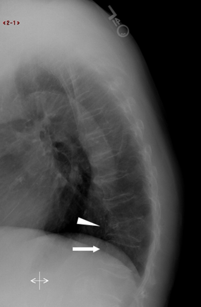
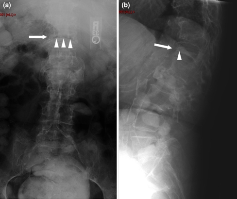
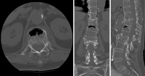
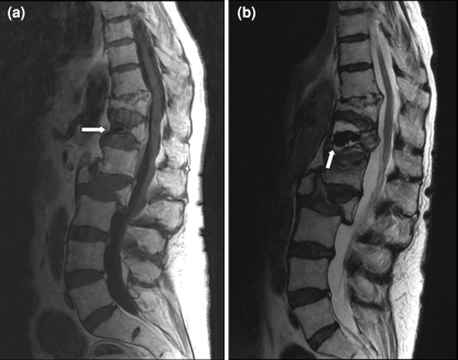
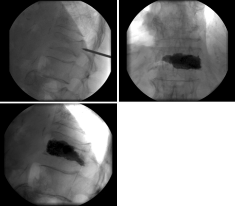
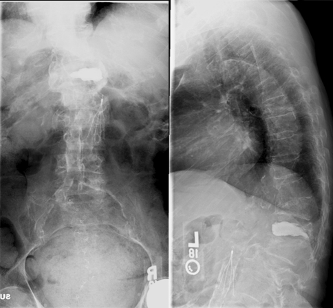
Delayed post-traumatic osteonecrosis, also known by its eponym Kummell's disease, is a rarely reported clinical entity that likely occurs with higher frequency than recognized. We highlight a case of a 75-year-old female household ambulator who presented with significant thoracolumbar pain and delayed T12 collapse after a ground-level fall. The patient had sustained a trivial fall at home 4 months prior to this presentation and had been hospitalized in our institution at that time for a general medical workup. Dedicated spine radiographs were not obtained during this visit. However, lateral chest radiograph demonstrated an intact T12 vertebral body. The patient was able to mobilize successfully with therapy and was discharged home. During the interim between the initial fall and subsequent presentation, she resumed physical activity including ambulating independently and performing various housework. Approximately 4 months following her initial injury, the patient returned to a local emergency department with vague complaints of abdominal pain without any history of recent fall or injury. After an unremarkable workup, the patient was sent home. Ten days later, she represented to our institution's emergency room with worsening pain. Radiographs and CT scan demonstrated interval collapse of the T12 vertebral body. A linear vacuum cleft was noted on X-rays and CT. An extensive workup to exclude other processes such as malignancy or infection, which was negative, ensued. Delayed post-traumatic vertebral collapse was diagnosed. A trial of medical management and therapy was attempted, but she continued to experience significant pain. A T12 vertebroplasty was therefore offered and performed to stabilize the injury and to relieve the pain. She was subsequently able to be discharged from the hospital and transitioned back to home life. At approximately 2 years following her injury, the patient was noted to be able to ambulate with a walking aid. Her final radiograph after her surgery demonstrated that the T12 vertebroplasty had maintained its height and sagittal alignment. This Grand Round case highlights the clinical presentation of Kummell's disease. Aspects of the clinical entity that will be discussed include a historical review of the disease, hallmark radiographic findings and treatment options.
Figures
Comment in
-
Expert's comment concerning Grand Rounds case entitled "Kümmell's disease: delayed post-traumatic osteonecrosis of the vertebral body" (by R. Ma, R. Chow, F. H. Shen).Eur Spine J. 2010 Jul;19(7):1071-2. doi: 10.1007/s00586-009-1204-5. Epub 2009 Nov 24. Eur Spine J. 2010. PMID: 19937066 Free PMC article. No abstract available.
References
-
- Kummell H. Die rarefizierende Ostitis der Wirbelkörper. Deutsche Med. 1895;21:180–181. doi: 10.1055/s-0029-1199707. - DOI
-
- Young WF, Brown D, Kendler A, Clements D. Delayed post-traumatic osteonecrosis of a vertebral body (Kummell’s disease) Acta Orthop Belg. 2002;68(1):13–19. - PubMed
-
- Chou LH, Knight RQ. Idiopathic avascular necrosis of a vertebral body. Case report and literature review. Spine (Phila Pa 1976) 1997;22(16):1928–1932. - PubMed
-
- Swartz K, Fee D. Kummell’s disease: a case report and literature review. Spine (Phila Pa 1976) 2008;33(5):E152–E155. - PubMed
Publication types
MeSH terms
LinkOut - more resources
Full Text Sources
Medical
