. 2010 Jan;88(1):47-60.
doi: 10.1007/s00109-009-0524-6. Epub 2009 Dec 1.

The inhibition of pyruvate dehydrogenase kinase improves impaired cardiac function and electrical remodeling in two models of right ventricular hypertrophy: resuscitating the hibernating right ventricle

Affiliations

The inhibition of pyruvate dehydrogenase kinase improves impaired cardiac function and electrical remodeling in two models of right ventricular hypertrophy: resuscitating the hibernating right ventricle

Lin Piao et al. J Mol Med (Berl). 2010 Jan.

Abstract

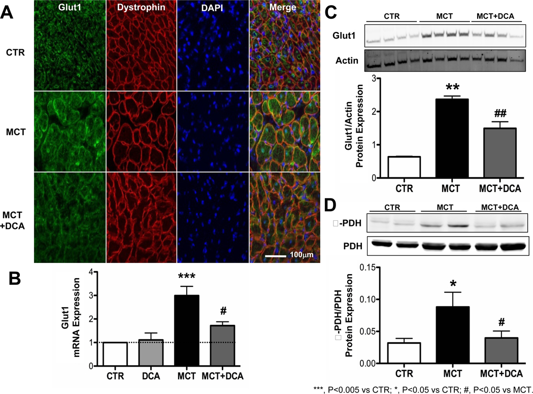
Right ventricular hypertrophy (RVH) and RV failure contribute to morbidity and mortality in pulmonary arterial hypertension (PAH). The cause of RV dysfunction and the feasibility of therapeutically targeting the RV are uncertain. We hypothesized that RV dysfunction and electrical remodeling in RVH result, in part, from a glycolytic shift in the myocyte, caused by activation of pyruvate dehydrogenase kinase (PDK). We studied two complementary rat models: RVH + PAH (induced by monocrotaline) and RVH + without PAH (induced by pulmonary artery banding (PAB)). Monocrotaline RVH reduced RV O(2)-consumption and enhanced glycolysis. RV 2-fluoro-2-deoxy-glucose uptake, Glut-1 expression, and pyruvate dehydrogenase phosphorylation increased in monocrotaline RVH. The RV monophasic action potential duration and QT(c) interval were prolonged due to decreased expression of repolarizing voltage-gated K(+) channels (Kv1.5, Kv4.2). In the RV working heart model, the PDK inhibitor, dichloroacetate, acutely increased glucose oxidation and cardiac work in monocrotaline RVH. Chronic dichloroacetate therapy improved RV repolarization and RV function in vivo and in the RV Langendorff model. In PAB-induced RVH, a similar reduction in cardiac output and glycolytic shift occurred and it too improved with dichloroacetate. In PAB-RVH, the benefit of dichloroacetate on cardiac output was approximately 1/3 that in monocrotaline RVH. The larger effects in monocrotaline RVH likely reflect dichloroacetate's dual metabolic benefits in that model: regression of vascular disease and direct effects on the RV. Reduction in RV function and electrical remodeling in two models of RVH relevant to human disease (PAH and pulmonic stenosis) result, in part, from a PDK-mediated glycolytic shift in the RV. PDK inhibition partially restores RV function and regresses RVH by restoring RV repolarization and enhancing glucose oxidation. Recognition that a PDK-mediated metabolic shift contributes to contractile and ionic dysfunction in RVH offers insight into the pathophysiology and treatment of RVH.


Conflict of interest statement

DISCLOSURES

The authors have no conflicts to disclose.

Figures

Figure 1
Figure 1. Dichloroacetate reduces RVH caused by Monocrotaline and improves cardiac function
The upper panel shows the chronic regression protocol of the administration of monocrotaline and dichloroacetate. A. Representative and mean data showing that dichloroacetate (DCA) lengthens PAAT (lowers PAP) in monocrotaline-induced RVH. B. The RVH is reduced by dichloroacetate. C. H&E staining showing that dichloroacetate decreases the hypertrophy of RV myocytes. D. Increased right ventricular free wall (RVFW) thickness is reduced by DCA. E. The impaired RVFW systolic thickening in RVH is increased by DCA. F. Representative traces and mean values showing dichloroacetate reduces RVSP in RVH in the RV Langendorff model. G. DCA restores cardiac output in vivo in the MCT+DCA group. CTR, Control group; DCA, Dichloroacetate group; MCT, Monocrotaline group; MCT+DCA, Monocrotaline+Dichloroacetate group.
Figure 2
Figure 2. Dichloroacetate acutely improves cardiac function and increases glucose oxidation in RVH in a working heart model
A Cardiac work increased 40 minutes after addition of DCA to the perfusate, only in the RVH group. B. Addition of 1 mM DCA to the perfusate increased glucose oxidation 4-fold in monocrotaline hearts. C. Glycolysis increases in RVH.
Figure 3
Figure 3. Reduced O2-consumption and increased FDG uptake in RVH
A and B. Representative traces and mean data showing DCA reverses the depressed O2-consumption in RVH. C and D. Representative images and mean data showing increased FDG uptake in the RV in RVH on PET scans.
Figure 4
Figure 4. Increased Glut1 expression and PDH phosphorylation in RVH are reduced by dichloroacetate
A. Immunostaining showing that the increased Glut1 expression in RV myocytes in RVH is reduced by DCA. The merge of the staining of Glut1 (green) and dystrophin (red) in RV shows that more Glut1 is expressed at the myocyte membrane in RVH. B and C, Glut1 mRNA and protein levels are significantly increased in RVH and DCA normalizes expression. D. Increased phosphorylation of PDH in RVH is reduced by dichloroacetate.
Figure 5
Figure 5. Dichloroacetate shortens prolonged MAP duration and QTc in RVH
A, B and C, Representative traces and mean data showing MAPD20 and MAPD90 are significantly prolonged in the MCT group vs CTR and that repolarization is improved by DCA. D and E, Representative traces and mean data of lead II surface ECG. DCA reduces the QTc prolongation in RVH. Red highlighted arrows indicate QTc intervals.
Figure 6
Figure 6. Repolarizing Kv channels that are downregulated in RVH are partially restored by dichloroacetate
A, B and C, Kv1.2, Kv1.5 and Kv4.2 are significantly downregulated in RVH and DCA partially reverses this downregulation. D, E and F, Immunoblotting showing the downregulation of Kv1.2, Kv1.5 and Kv4.2 by monocrotaline. Kv1.5 expression in RH is enhanced by DCA.
Figure 7
Figure 7. Dichloroacetate reverses RVH induced by pulmonary artery banding and improves cardiac function
A. H&E staining and mean data of cell size show DCA reduces cell size in RVH. B representative traces showing the gradient across the stenosis in the PAB model. Mean±SEM of hemodynamic data in this model. C. The reduced RVFW systolic thickening in PAB is improved by DCA. D. Reduced cardiac output, measured by thermodilution, is improved by DCA.
Figure 8
Figure 8. Dichloroacetate reverses metabolic changes in RVH induced by pulmonary artery banding
A. Immunostaining of Glut1 shows that the increased Glut1 expression at myocytes membranes is reduced by DCA. B. DCA reverses the impaired oxygen consumption in RVH. C. Dichloroacetate causes a greater relative increase in CO in the Monocrotaline versus the PAB Model. D. Proposed mechanism of hibernating RV myocardium in RVH. The activation of PDK in RVH reduces the coupling of glycolysis to glucose oxidation. These metabolic changes are (in part) rapidly reversible by dichloroacetate, which improves contractility by restoring normal RV energetics. PDK activation also inhibits the expression of Kv channels, which prolongs APD. Prolonged APD may also contribute to impairment of RV function in RVH.

Similar articles

Cited by

References

    1. Bonnet S, Michelakis ED, Porter CJ, Andrade-Navarro MA, Thebaud B, Haromy A, Harry G, Moudgil R, McMurtry MS, Weir EK, Archer SL. An abnormal mitochondrial-hypoxia inducible factor-1alpha-Kv channel pathway disrupts oxygen sensing and triggers pulmonary arterial hypertension in fawn hooded rats: similarities to human pulmonary arterial hypertension. Circulation. 2006;113:2630–2641. - PubMed
    1. van Wolferen SA, Marcus JT, Boonstra A, Marques KM, Bronzwaer JG, Spreeuwenberg MD, Postmus PE, Vonk-Noordegraaf A. Prognostic value of right ventricular mass, volume, and function in idiopathic pulmonary arterial hypertension. Eur Heart J. 2007;28:1250–1257. - PubMed
    1. Hessel MH, Steendijk P, den Adel B, Schutte CI, van der Laarse A. Characterization of right ventricular function after monocrotaline-induced pulmonary hypertension in the intact rat. Am J Physiol Heart Circ Physiol. 2006;291:H2424–H2430. - PubMed
    1. Lamberts RR, Caldenhoven E, Lansink M, Witte G, Vaessen RJ, St Cyr JA, Stienen GJ. Preservation of diastolic function in monocrotaline-induced right ventricular hypertrophy in rats. Am J Physiol Heart Circ Physiol. 2007;293:H1869–H1876. - PubMed
    1. Lee JK, Kodama I, Honjo H, Anno T, Kamiya K, Toyama J. Stage-dependent changes in membrane currents in rats with monocrotaline-induced right ventricular hypertrophy. Am J Physiol. 1997;272:H2833–H2842. - PubMed

Publication types

MeSH terms

Substances

LinkOut - more resources