

Differentiation dependent expression of urocortin's mRNA and peptide in human osteoprogenitor cells: influence of BMP-2, TGF-beta-1 and dexamethasone
- PMID: 19949969
- PMCID: PMC2834774
- DOI: 10.1007/s10735-009-9244-z
Differentiation dependent expression of urocortin's mRNA and peptide in human osteoprogenitor cells: influence of BMP-2, TGF-beta-1 and dexamethasone
Abstract
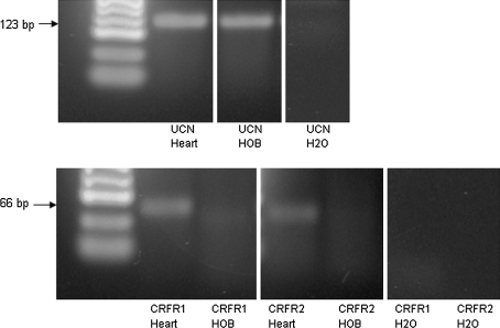
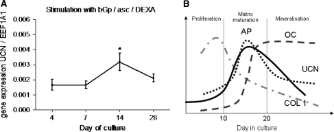
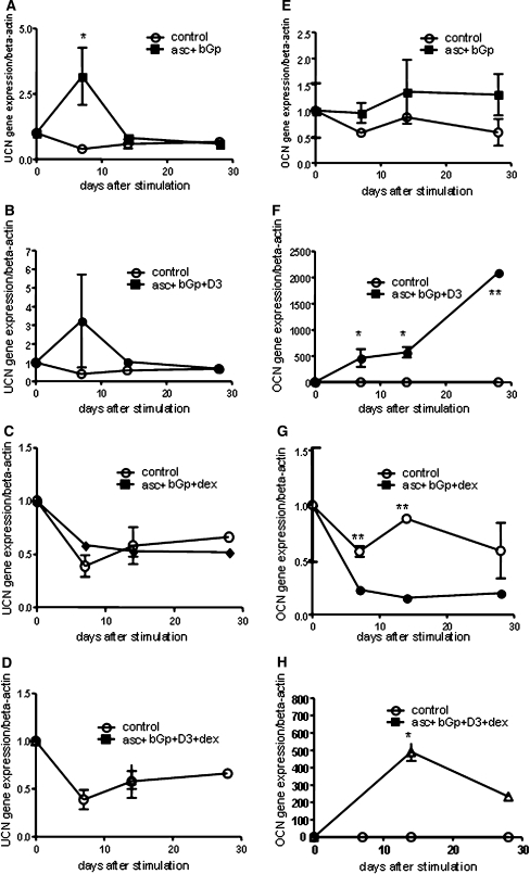
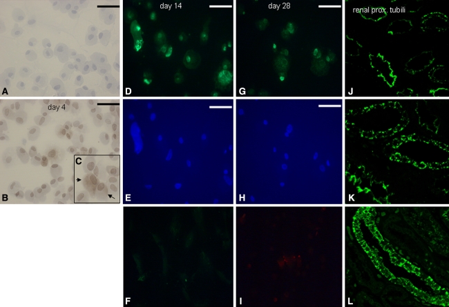
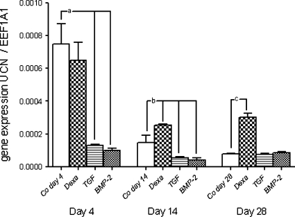
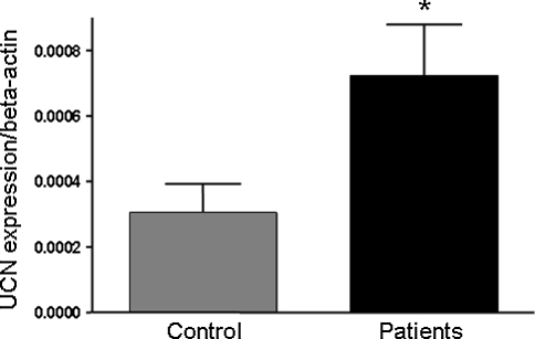
Urocortin-1 (UCN) a corticotropin releasing-factor (CRF) related peptide, has been found to be expressed in many different tissues like the central nervous system, the cardiovascular system, adipose tissue, and skeletal muscle. The effects of UCN are mediated via stimulation of CRF-receptors 1 and 2 (CRFR1 and 2, CRFR's) with a high affinity for CRFR2. It has been shown that the CRF-related peptides and CRFR's are involved in the regulation of stress-related endocrine, autonomic and behavioural responses. Using immunocytochemistry, immunohistochemistry and RT-PCR, we now can show the differentiation dependent expression of UCN mRNA and peptide in human mesenchymal progenitor cells (MSCs) directed to the osteoblastic phenotype for the first time. UCN expression was down regulated by TGF-beta and BMP-2 in the early proliferation phase of osteoblast development, whereas dexamethasone (dex) minimally induced UCN gene expression during matrix maturation after 24 h stimulation. Stimulation of MSCs for 28 days with ascorbate/beta-glycerophosphate (asc/bGp) induced UCN gene expression at day 14. This effect was prevented when using 1,25-vitamin D3 or dex in addition. There was no obvious correlation to osteocalcin (OCN) gene expression in these experiments. In MSCs from patients with metabolic bone disease (n = 9) UCN gene expression was significantly higher compared to MSCs from normal controls (n = 6). Human MSCs did not express any of the CRFR's during differentiation to osteoblasts. Our results indicate that UCN is produced during the development of MSCs to osteoblasts and differentially regulated during culture as well as by differentiation factors. The expression is maximal between proliferation and matrix maturation phase. However, UCN does not seem to act on the osteoblast itself as shown by the missing CRFR's. Our results suggest new perspectives on the role of urocortin in human skeletal tissue in health and disease.
Figures
Similar articles
-
Differential regulation of Cbfa1/Runx2 and osteocalcin gene expression by vitamin-D3, dexamethasone, and local growth factors in primary human osteoblasts.J Cell Biochem. 2002;86(2):348-56. doi: 10.1002/jcb.10220. J Cell Biochem. 2002. PMID: 12112004
-
Urocortin II gene is highly expressed in mouse skin and skeletal muscle tissues: localization, basal expression in corticotropin-releasing factor receptor (CRFR) 1- and CRFR2-null mice, and regulation by glucocorticoids.Endocrinology. 2004 May;145(5):2445-57. doi: 10.1210/en.2003-1570. Epub 2004 Jan 21. Endocrinology. 2004. PMID: 14736736
-
Dexamethasone modulates BMP-2 effects on mesenchymal stem cells in vitro.J Orthop Res. 2008 Nov;26(11):1440-8. doi: 10.1002/jor.20565. J Orthop Res. 2008. PMID: 18404732
-
Differential effects of dexamethasone on the chondrogenesis of mesenchymal stromal cells: influence of microenvironment, tissue origin and growth factor.Eur Cell Mater. 2011 Nov 24;22:302-19; discussion 319-20. doi: 10.22203/ecm.v022a23. Eur Cell Mater. 2011. PMID: 22116649
-
Urocortin and corticotropin-releasing factor receptor 2 in human renal cell carcinoma: disruption of an endogenous inhibitor of angiogenesis and proliferation.World J Urol. 2009 Dec;27(6):825-30. doi: 10.1007/s00345-009-0417-x. World J Urol. 2009. PMID: 19437022 Free PMC article.
Cited by
-
Dose dependent effect of C-type natriuretic peptide signaling in glycosaminoglycan synthesis during TGF-β1 induced chondrogenic differentiation of mesenchymal stem cells.J Mol Histol. 2010 Oct;41(4-5):247-58. doi: 10.1007/s10735-010-9284-4. Epub 2010 Aug 19. J Mol Histol. 2010. PMID: 20721606
-
Bone morphogenetic protein signaling: implications in urology.Korean J Urol. 2010 Aug;51(8):511-7. doi: 10.4111/kju.2010.51.8.511. Epub 2010 Aug 18. Korean J Urol. 2010. PMID: 20733955 Free PMC article.
-
Effect of Urocortin on strength and microarchitecture of osteopenic rat femur.J Bone Miner Metab. 2015 Mar;33(2):154-60. doi: 10.1007/s00774-014-0578-6. Epub 2014 Mar 16. J Bone Miner Metab. 2015. PMID: 24633537
References
MeSH terms
Substances
LinkOut - more resources
Full Text Sources
Miscellaneous
