. 2010 Jun;12(3):349-58.
doi: 10.1007/s11307-009-0270-8. Epub 2009 Dec 1.

Molecular imaging of the translocator protein (TSPO) in a pre-clinical model of breast cancer

Affiliations

Molecular imaging of the translocator protein (TSPO) in a pre-clinical model of breast cancer

Shelby K Wyatt et al. Mol Imaging Biol. 2010 Jun.

Abstract

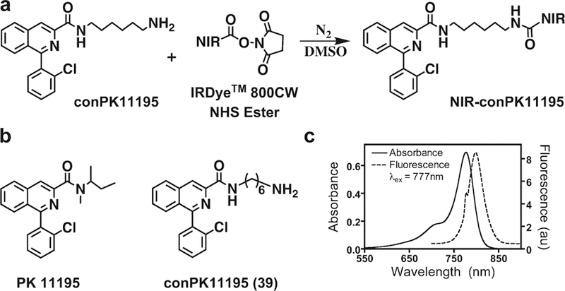
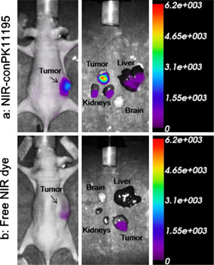
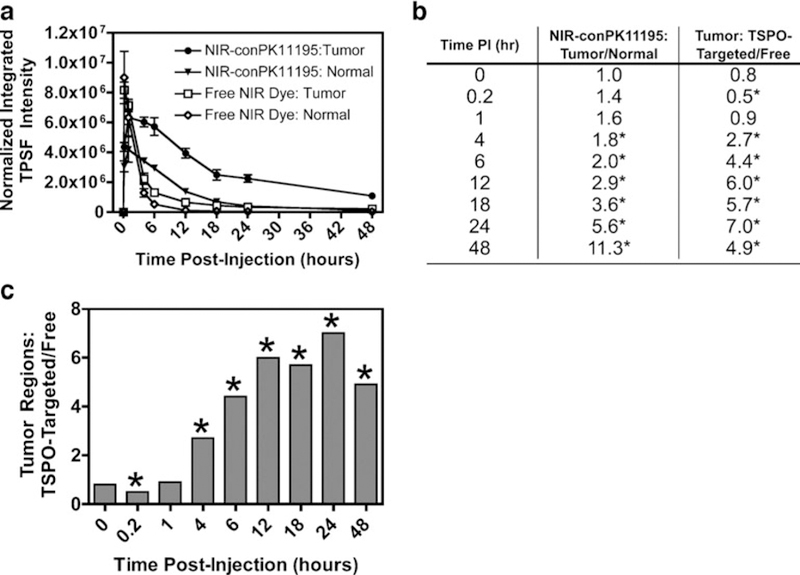
Purpose: To quantitatively evaluate the utility of a translocator protein (TSPO)-targeted near-infrared (NIR) probe (NIR-conPK11195) for in vivo molecular imaging of TSPO in breast cancer.

Procedures: NIR-conPK11195 uptake and TSPO-specificity were validated in TSPO-expressing human breast adenocarcinoma cells (MDA-MB-231). In vivo NIR-conPK11195 biodistribution and accumulation were quantitatively evaluated in athymic nude mice bearing MDA-MB-231 xenografts.

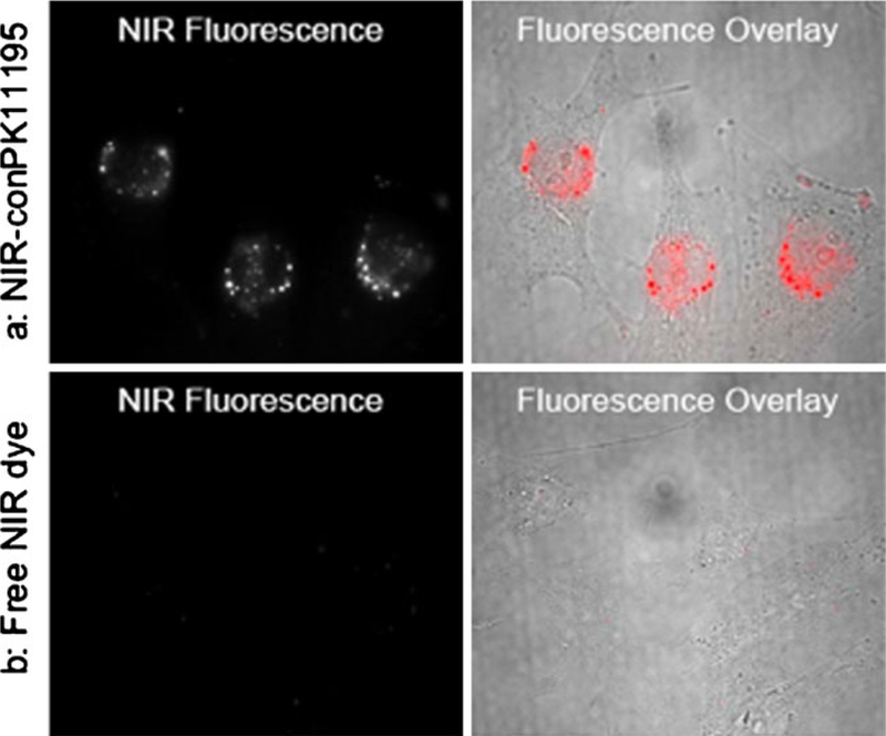
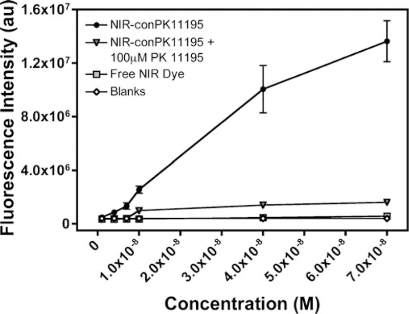
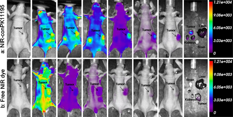
Results: Fluorescence micrographs illustrated intracellular labeling of MDA-MB-231 cells by NIR-conPK11195. Quantitative uptake and competition assays demonstrated dose-dependent (p < 0.001) and TSPO-specific (p < 0.001) NIR-conPK11195 uptake. In vivo, NIR-conPK11195 preferentially labeled MDA-MB-231 tumors with an 11-fold (p < 0.001) and 7-fold (p < 0.001) contrast enhancement over normal tissue and unconjugated NIR dye, respectively.

Conclusions: NIR-conPK11195 appears to be a promising TSPO-targeted molecular imaging agent for visualization and quantification of breast cancer cells in vivo. This research represents the first study to demonstrate the feasibility of TSPO imaging as an alternative breast cancer imaging approach.


Figures

Fig. 1
Fig. 1
a Synthetic pathway, b relevant chemical structures, and c spectroscopic properties of NIR-conPK11195 in CMF-PBS (λex =777 nm).
Fig. 2
Fig. 2
Live cell fluorescence microscopy illustrates appreciable NIR fluorescence and cellular uptake of NIR-conPK11195 (a), but not free NIR dye (b). MDA-MB-231 cells were incubated with 10 μM NIR-conPK11195 (a) or free NIR dye (b) for 30 min at 37°C in FBS-free medium. Pseudocolored fluorescence images overlaid onto the corresponding white light images also provide evidence of nuclear and perinuclear localization of NIR-conPK11195.
Fig. 3
Fig. 3
Cellular uptake and competition assays indicate significant dose dependent (p<0.001) and TSPO-specific (p<0.001) cellular uptake of NIR-conPK11195. The average fluorescence intensity per well±standard deviation was plotted as a function of NIR-conPK11195 concentration (unchallenged (circle) or in the presence of 100 μM PK 11195 (inverted triangle)) or free NIR dye concentration (square); undosed cells (diamond) serve as a negative control for autofluorescence. n=3 wells.
Fig. 4
Fig. 4
Direct comparison of the biodistribution and accumulation of the TSPO-targeted NIR-conPK11195 (a) and free NIR dye (b) in tumor-bearing mice demonstrates significantly different clearance profiles and enhanced preferential labeling of MDA-MB-231 tumors in vivo by NIR-conPK11195. Fluorescence images were normalized to laser power and integration time, overlaid onto the corresponding white light images, and displayed in terms of normalized photon counts over 48 h post-injection (pi). Images are representative of n=3 mice per group. Note: These images are all displayed on the same scale (0 to 12,149 counts) pre-injection, 1, 4, 6, 12, 24, and 48 h pi (left to right).
Fig. 5
Fig. 5
NIR-conPK11195 preferentially labels MDA-MB-231 tumors in vivo and substantially enhances the tumor-associated signal over free NIR dye (see also Fig. 6). a and b Fluorescence overlays of MDA-MB-231 tumor-bearing mice and harvested tissues ∼48 h pi of NIR-conPK11195 and free NIR dye, respectively. Representative of n=3 mice per group.
Fig. 6
Fig. 6
a Time-activity curves illustrate the distinct clearance profile of NIR-conPK11195 relative to free NIR dye, further demonstrate tumor specificity of NIR-conPK11195 in vivo, and reveal the significant signal enhancement resulting from the TSPO-targeted NIR molecular imaging probe. b ROI contrast ratios. c Graphical representation of the TSPO-targeted to free NIR dye ratio demonstrating significant fluorescence signal enhancement from the NIR-conPK11195 relative to the free NIR dye. *p<0.001.

Similar articles

Cited by

References

    1. American Cancer Society (2009) Cancer Facts and Figures Atlanta: American Cancer Society
    1. Elmore JG, Armstrong K, Lehman CD, Fletcher SW (2005) Screening for breast cancer. JAMA 293(10):1245–1256 - PMC - PubMed
    1. Carney PA, Miglioretti DL, Yankaskas BC et al. (2003) Individual and combined effects of age, breast density, and hormone replacement therapy use on the accuracy of screening mammography. Ann Intern Med 138(3):168–175 - PubMed
    1. DeMartini W, Lehman C, Partridge S (2008) Breast MRI for cancer detection and characterization: a review of evidence-based clinical applications. Acad Radiol 15(4):408–416 - PubMed
    1. Stavros AT, Thickman D, Rapp CL, Dennis MA, Parker SH, Sisney GA (1995) Solid breast nodules: use of sonography to distinguish between benign and malignant lesions. Radiology 196(1):123–134 - PubMed

Publication types