. 2010 Feb;40(2):449-59.
doi: 10.1002/eji.200939586.

Adenosine mediated desensitization of cAMP signaling enhances T-cell responses

Affiliations

Adenosine mediated desensitization of cAMP signaling enhances T-cell responses

Ailian Yang et al. Eur J Immunol. 2010 Feb.

Abstract

Adenosine has long been regarded as a crucial anti-inflammatory agent that protects the host from excessive damage. It has been reported to play an important role in suppressing immune activation, particularly that of T cells. However, it is a general observation that induction of T-cell activation is an efficient event despite the high adenosine levels that are often present in the affected host due to injury or stress. We report here that prior to antigenic stimulation via TCR/CD3, exposure of T cells to adenosine desensitizes adenosine receptors, so as to create a window of time where the T cells are insensitive to this ubiquitous suppressor. T cells from mice that were pre-exposed to this manipulation showed stronger responses to antigenic stimulation; therefore, the P1 adenosine receptor desensitization demonstrated an adjuvant-like effect. Our results suggest that adenosine receptor desensitization may be a mechanism for T cells to escape the general suppression during early points of T-cell activation and may emerge as a potential alternative for vaccine adjuvants.


Figures
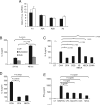
Figure 1
Figure 1
Adenosine suppresses CD8+ and CD4+ T-cell activation. (A) Real-time PCR ΔCT analysis of P1R messages versus GAPDH. Filled bars: purified splenic CD4+ cells from untreated C57BL/6 mice; Open bars: from C57BL/6 mice pre-injected with NECA. (B) CD4+ T cells from C57BL/6 mice were purified and incubated with plate-bound anti-CD3 (activation) mAb in 96-well plates, with or without 5 μM adenosine as indicated. Supernatants were collected after different time points and IL-2 levels were determined by an ELISA kit. All data points shown (and henceforth) were performed in triplicates. UT: untreated. (C) Similar to (B), splenocytes (2 × 10⁶/mL) from OT-II mice were activated with a soluble peptide ISQAVHAAHAEINEAGR (OVA 323–339) (1 mg/mL) for IL-2 production in the presence of adenosine receptor agonists. CPA (10 μM): A1 agonist; CGS (10 μM): A2A agonist; IB-MECA (10 μM): A3 agonist, NECA (10 μM): a non-selective adenosine agonist, EHNA (10 μM): an ADA inhibitor. (D) CD4+ T cells from C57BL/6 mice were purified and incubated with anti-CD3 antibody as in (B), with or without adenosine agonist CPA (10 μM) or NECA (10 μM). Supernatants were collected at 24 h and IL-2 production was determined by ELISA. (E) Splenocytes (2 × 10⁶/mL) from OT-I mice were incubated with SIINFEKL (10–7M) with or without adenosine agonists as indicated. Supernatants were collected after 48 h and IFN-γ levels were measured with an ELISA kit.
Figure 2
Figure 2
Pre-exposure to adenosine leads to enhanced subsequent T-cell activation. (A) C57BL/6 mice were injected i.p. with PBS, CPA (20 μM/kg), CGS (13 μM/kg) or NECA (1 μM/kg) (50 μL total volume). The same treatment was repeated after 18 h. After 24 h, mice were euthanized and splenocytes were harvested. CD4+ T cells were purified and activated with anti-CD3 antibody. IL-2 production was measured after 24 h. (B) same as A except CPA was co-injected with DPCPX, and supernatants were collected both at 24 and 48 h. (C) Same as (A), except CD8+ T cells were purified and tested. (D) Splenocytes from OT-I or OT-II were incubated with or without CPA (10 μM) or NECA (10 μM) for 24 h and were then stained with Annexin V and Propidium Iodide. Cells were analyzed by FACS to determine their viability. The percentage of apoptotic cells is indicated in each panel.
Figure 3
Figure 3
The enhanced T-cell response is mediated by NECA and CPA. (A) PCR genotyping of C57BL/6 and A1R KO mice for A1R deletion was performed as described in Materials and methods. Only WT and deletion mutant gene products were present in C57BL/6 and A1R KO mice, respectively. (B) cDNA, reverse transcribed from mRNA isolated from kidney cell lysate from C57BL/6 and A1R KO mice, were analyzed by PCR for the presence of A1R transcript as described in Materials and methods. Rodent GAPDH was used as the control. GAPDH transcript controls were present in both preparations. (C) A1R KO mice were injected i.p. with PBS or CPA (20 μM/kg) in a total volume of 50 μL. The same treatment was repeated after 18 h. After 24 h, mice were euthanized and splenocytes were harvested. CD4+ T cells were purified and activated with anti-CD3 antibody. IL-2 production was measured after 24 h incubation. (D) Similar to Fig. 2A except that A2AR KO mice were used in place. (E) CD4+ T cells from A1R KO mice were purified and incubated with anti-CD3 antibody, with or without adenosine agonist CPA (10 μM) or NECA (10 μM). IL-2 in the culture supernatants was measured after 24 h.
Figure 4
Figure 4
Treatments leading to increased T-cell responses upregulate cAMP production. (A) and (B) Purified C57BL/6 CD4+ T cells from C57BL/6 were suspended in the cell culture medium and stimulated with immobilized anti-CD3 antibody in the presence of adenosine agonists or Forskolin. At different time points, media were aspirated and lysed with 0.1 M HCL (20 μL in 200 μmL medium), and then incubated at room temperature for 20 min. Supernatants after centrifugation were acetylated with KOH and acetic anhydride. The concentration of cAMP was derived from the raw data following manufacturer's instructions. (A) The preliminary experiment that determined the optimal time point for (B). UT: untreated; For or F: Forskolin; C: CPA; A3ag or A3: IB-MECA. (C) Purified CD4+ T cells were suspended in the cell culture medium and incubated on immobilized anti-CD3 in the presence of adenosine agonist CPA (10 μM) or CGS (10 μM), with or without antibody CT (10 ng/mL) or PT (2 ng/mL). Supernatants were collected after 24 h incubation and IL-2 was determined by an ELISA kit. CON: anti-CD3 antibody only.
Figure 5
Figure 5
Pre-exposure to adenosine leads to P1R desensitization. (A) C57BL/6 mice were injected i.p. with PBS or NECA (1 μM/kg). 18 h later, the same treatment was repeated. After 24 h, mice were euthanized and splenocytes were harvested. CD4+ T cells were purified and activated with anti-CD3 antibody with or without the presence of NECA (10 μM). IL-2 levels in the overnight supernatant were determined by ELISA. (B) Splenocytes from C57BL/6 were incubated with or without NECA (10 μM) overnight. Splenocytes were washed, and CD4+ T cells from C57BL/6 were purified and activated with anti-CD3 antibody. The rest of the assay was identical to (A). (C) C57BL/6 mice were injected i.p. with PBS or NECA (1 μM/kg). The same treatment was repeated after 18 h. After 24 h, mice were euthanized and splenocytes were harvested. CD4+ T cells were purified and activated with anti-CD3 antibody in the absence or presence of NECA (10 μM). After a 15 min incubation, cAMP was determined as in Fig. 4A and B. The top X-axis labels are the pre-treatment in vivo and the bottom labels are the subsequent in vitro treatment.
Figure 6
Figure 6
Desensitization of P1R is rapidly reversible. C57BL/6 mice were pre-injected i.p. with CPA as in Fig. 2A. After 24 h, mice were sacrificed and CD4+ T cells were purified from the splenocytes and stimulated with plate-bound anti-CD3 antibody. NECA (1 μM) was added at the beginning of or at various points into the culture. The IL-2 production in the supernatant was measured after 24 h.
Figure 7
Figure 7
The transient desensitization of P1R exerts an adjuvant effect on T-cell activation. (A) C57BL/6 mice were injected i.p. with PBS or CPA (20 μM/kg). The same treatment was repeated after 18 h. After 24 h, soluble OVA (200 mg/kg) was used to immunize mice as described in Materials and methods. As a control, a mixture of CFA+OVA was injected into control mice. After 7 days, mice were euthanized and splenocytes were harvested and activated with soluble OVA. IL-2 concentration in the supernatant was determined by ELISA. (B) and (C) OT-I mice were injected i.p. with PBS, CPA or NECA as in (A). After 24 h, latex beads (5 μg) were injected s.c. into mice. As a control, a mixture of CFA+latex beads was injected into control mice. In total 24 h later, mice were euthanized and splenocytes (10 × 106/mL for (B) or 5 × 10⁶/mL for (C)) were harvested and activated with SIINFEKL. After a 24-h incubation, IL-2 in the supernatant was determined by ELISA. This figure was independently repeated four times.

Similar articles

Cited by

References

    1. Hasko G, Cronstein BN. Adenosine: an endogenous regulator of innate immunity. Trends Immunol. 2004;25:33–39. - PubMed
    1. Hasko G, Pacher P, Vizi ES, Illes P. Adenosine receptor signaling in the brain immune system. Trends Pharmacol. Sci. 2005;26:511–516. - PMC - PubMed
    1. Hershfield MS. New insights into adenosine-receptor-mediated immunosuppression and the role of adenosine in causing the immunodeficiency associated with adenosine deaminase deficiency. Eur. J. Immunol. 2005;35:25–30. - PubMed
    1. Yaar R, Jones MR, Chen JF, Ravid K. Animal models for the study of adenosine receptor function. J. Cell. Physiol. 2005;202:9–20. - PubMed
    1. Ohta A, Sitkovsky M. Role of G-protein-coupled adenosine receptors in downregulation of inflammation and protection from tissue damage. Nature. 2001;414:916–920. - PubMed

Publication types

MeSH terms

LinkOut - more resources