. 2010 Mar;40(3):764-9.
doi: 10.1002/eji.200939996.

Experimental cerebral malaria progresses independently of the Nlrp3 inflammasome

Affiliations

Experimental cerebral malaria progresses independently of the Nlrp3 inflammasome

Thornik Reimer et al. Eur J Immunol. 2010 Mar.

Abstract

Cerebral malaria is the most severe complication of Plasmodium falciparum infection in humans and the pathogenesis is still unclear. Using the P. berghei ANKA infection model of mice, we investigated a potential involvement of Nlrp3 and the inflammasome in the pathogenesis of cerebral malaria. Nlrp3 mRNA expression was upregulated in brain endothelial cells after exposure to P. berghei ANKA. Although beta-hematin, a synthetic compound of the parasites heme polymer hemozoin, induced the release of IL-1beta in macrophages through Nlrp3, we did not obtain evidence for a role of IL-1beta in vivo. Nlrp3 knock-out mice displayed a delayed onset of cerebral malaria; however, mice deficient in caspase-1, the adaptor protein ASC or the IL-1 receptor succumbed as WT mice. These results indicate that the role of Nlrp3 in experimental cerebral malaria is independent of the inflammasome and the IL-1 receptor pathway.


Conflict of interest statement

Conflict of interest

The authors declare no financial or commercial conflict of interest.

Figures

Figure 1
Figure 1. The role of the Nlrp3 inflammasome in response to beta-hematin and Plasmodium extracts
Macrophages from WT or Nlrp3−/− mice were incubated in the presence of 1 ug/ml LPS, 250 µg/ml beta-hematin (b-HT) or 1 mg/ml Plasmodium falciparum 3D7 extracts (PfCE). Where indicated, either 1 ug/ml LPS or 5 mM ATP was added to the beta-hematin and Plasmodium falciparum culture respectively. LPS was added for the entire incubation period whereas ATP was only added for the last 30 minutes. After 16 hours, culture supernatants were collected and IL-1β and IL-6 release were assessed by ELISA. Data show mean + SEM and are representative of 3 independent experiments.
Figure 2
Figure 2. Nlrp3 functions independently of the inflammasome in experimental cerebral malaria
Wild-type, Nlrp3−/−, P2X7R−/−, ASC−/−, Caspase-1−/− and IL-1R−/− mice were intraperitoneally infected with 106 Plasmodium berghei ANKA infected erythrocytes and monitored for survival. Ten mice per group were used for all groups, except for the P2X7 receptor mice. A representative survival pattern of at least two independent experiments is shown, except for the P2X7R−/− mice, which has only been performed once with five mice per group. Statistical differences were assessed using Gehan-Breslow-Wilcoxon Test and are indicated in the Figures.
Figure 3
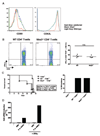
Figure 3. Nlrp3 does not function in T-cells but is induced in brain endothelial cells
Splenocytes were isolated from wild-type and Nlrp3−/− mice after 5 days of infection with Plasmodium berghei ANKA and stained for the expression of CD69, CD62L (A), and the expression of IFN-γ (B). Data are representative of three independent experiments. Data for individual mice are plotted as a dot plot. Statistical differences were assessed using the unpaired Student’s t-test. (C) Purified CD4+ and CD8+ T-cells from wild-type and Nlrp3−/− mice were adoptively transferred into TCRβ −/− recipients. 24 hours after transfer, recipients were infected with 1 × 10⁶ P. berghei ANKA infected red blood cells and monitored for survival (left panel) and incidence of cerebral malaria (right panel) at day 10. Statistical differences were assessed using Gehan-Breslow-Wilcoxon test and are as indicated. Data are representative of one experiments. (D) Mouse brain microvascular endothelial cells were incubated with uninfected erythrocytes or P. berghei ANKA parasitized erythrocytes for 16 hours. Where indicated, 100 ng/ml recombinant murine IFN-γ was added. RNA was collected, and Nlrp3 expression was assessed using quantitative PCR. Data are expressed as fold mRNA induction over untreated control samples (2−ΔΔCt). Data are representative of two independent experiments.

References

    1. Miller LH, Baruch DI, Marsh K, Doumbo OK. The pathogenic basis of malaria. Nature. 2002;415:673–679. - PubMed
    1. Schofield L, Grau GE. Immunological processes in malaria pathogenesis. Nat. Rev. Immunol. 2005;5:722–735. - PubMed
    1. Potter S, Chan-Ling T, Ball HJ, Mansour H, Mitchell A, Maluish L, Hunt NH. Perforin mediated apoptosis of cerebral microvascular endothelial cells during experimental cerebral malaria. Int. J. Parasitol. 2006;36:485–496. - PubMed
    1. Combes V, Rosenkranz AR, Redard M, Pizzolato G, Lepidi H, Vestweber D, Mayadas TN, Grau GE. Pathogenic Role of P-Selectin in Experimental Cerebral Malaria: Importance of the Endothelial Compartment. Am. J. Pathol. 2004;164:781–786. - PMC - PubMed
    1. Scholl PF, Tripathi AK, Sullivan DJ. Bioavailable iron and heme metabolism in Plasmodium falciparum. Curr. Top. Microbiol. Immunol. 2005;295:293–324. - PubMed

Publication types

MeSH terms