. 2009 Dec;60(12):3651-62.
doi: 10.1002/art.25006.

Inflammatory arthritis in caspase 1 gene-deficient mice: contribution of proteinase 3 to caspase 1-independent production of bioactive interleukin-1beta

Affiliations

Inflammatory arthritis in caspase 1 gene-deficient mice: contribution of proteinase 3 to caspase 1-independent production of bioactive interleukin-1beta

Leo A B Joosten et al. Arthritis Rheum. 2009 Dec.

Abstract

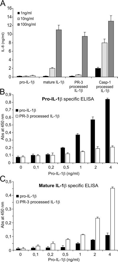
Objective: Caspase 1, a known cysteine protease, is a critical component of the inflammasome. Both caspase 1 and neutrophil serine proteases such as proteinase 3 (PR3) can process pro-interleukin-1beta (proIL-1beta), a crucial cytokine linked to the pathogenesis of rheumatoid arthritis. This study was undertaken to establish the relative importance of caspase 1 and serine proteases in mouse models of acute and chronic inflammatory arthritis.

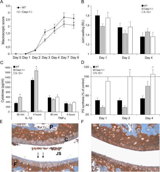
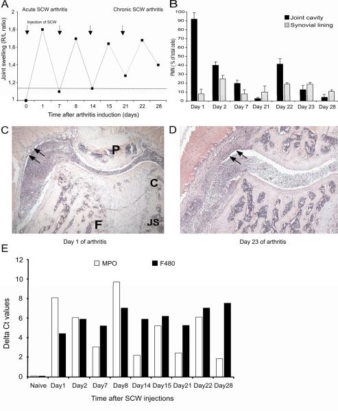
Methods: Acute and chronic arthritis were induced in caspase 1-/- mice, and the lack of caspase 1 was investigated for its effects on joint swelling, cartilage metabolism, and histopathologic features. In addition, caspase 1 activity was inhibited in mice lacking active cysteine proteases, and the effects of dual blockade of caspase 1 and serine proteases on arthritis severity and histopathologic features were evaluated.

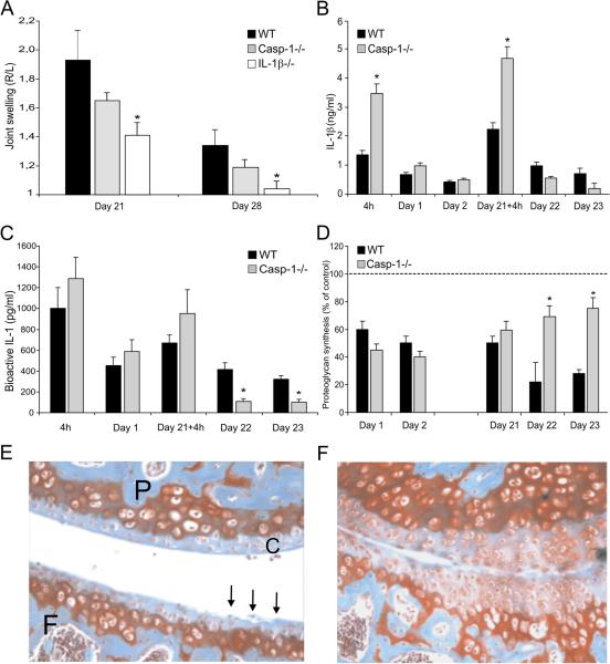
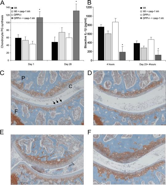
Results: Surprisingly, caspase 1-/- mice, in a model of acute (neutrophil-dominated) arthritis, developed joint swelling to an extent similar to that in wild-type control mice. Joint fluid concentrations of bioactive IL-1beta were comparable in caspase 1-/- mice and controls. In contrast, induction of chronic arthritis (characterized by minimal numbers of neutrophils) in caspase 1-/- mice led to reduced joint inflammation and less cartilage damage, implying a caspase 1-dependent role in this process. In mice lacking neutrophil serine PR3, inhibition of caspase 1 activity resulted in decreased bioactive IL-1beta concentrations in the synovial tissue and less suppression of chondrocyte anabolic function. In addition, dual blockade of both PR3 and caspase 1 led to protection against cartilage and bone destruction.

Conclusion: Caspase 1 deficiency does not affect neutrophil-dominated joint inflammation, whereas in chronic arthritis, the lack of caspase 1 results in reduced joint inflammation and cartilage destruction. These findings suggest that inhibitors of caspase 1 are not able to interfere with the whole spectrum of IL-1beta production, and therefore such inhibitors may be of therapeutic value only in inflammatory conditions in which limited numbers of neutrophils are present.


Figures

Figure 1
Figure 1. Acute arthritis in caspase-1−/− mice
A. K/BxN arthritis in caspase-1−/− mice. Arthritis was induced by i.p injection of 200μl arthritic serum at day 0. Data are expressed as mean±SEM of 7 mice per group. B. Acute SCW-induced arthritis in caspase-1−/− mice. Joint swelling at days 1, 2 and 4 after intraarticular injection of 25μg SCW fragments, measured by radioactive 99mTc-uptake method. The swelling is expressed as a right-left ratio and a ratio > 1.15 is indicated as inflammation. Data are expressed as mean±sem of 7 mice per group. C. Local IL-1β (both IL-1β and proIL-1β) and TNFα concentrations in synovial tissue explants, 90 minutes and 4 hours after induction of arthritis. Data represents the mean±SEM of 5 explants per group. D. Chondrocyte metabolic function determined in patellar cartilage explants by 35S-sulphate incorporation. Note almost complete protection against inhibition of chondrocyte PG synthesis in IL-1β−/− mice at day 1 and 2. Data represents the mean±SEM of 5 explants per group. E. Cartilage proteoglycan (PG) depletion visualized by Safranin O staining (arrows indicated PG depletion) at day 7 of SCW arthritis, 200 × magnifications. P= patella, F= femur, C = cartilage, JS = joint space. F. Cartilage PG depletion in caspase-1−/−mice, noted the reduction of PG loss. *P<0.01 compared to wild-type control mice, Mann-Whitney U-test.
Figure 2
Figure 2. Induction of chronic SCW arthritis in C57Bl/6 mice
A: Arthritis, induced by intraarticular (i.a.) injections of 25 μg SCW fragments at days 0, 7, 14 and 21, leads to a chronic inflamed joint at day 28. Note that the joint swelling remains detectable after the second i.a. injection (dotted line indicates detectable joint swelling). B. Cellular influx during the course of chronic SCW-induced arthritis. Predominantly PMN influx in acute (1st injection) SCW- and shortly after the 4th reactivation (day 22) of chronic SCW-induced arthritis. C. PMN influx in the joint cavity at day 1 of SCW arthritis (wild-type mouse). H&E staining, 40×. D, Cellular influx in synovial lining and joint cavity at day 23 of chronic SCW arthritis. H&E staining, 40×. Note the difference in synovial lining between day1 and day 23 (arrows). E, Cell marker expression in inflamed synovium. MPO (PMN) and F4/80 (Macrophage) mRNA expression was determine during the course of SCW-induced arthritis. Note that MPO is predominantly expressed in the acute stages whereas F4/80 is seen at the more chronic stages. For details, see Figure 1.
Figure 3
Figure 3. Chronic SCW-induced arthritis in caspase-1−/− mice
A. Joint swelling at days 21 and 28 in caspase-1−/− mice, compared to wild-type and IL-1β−/− mice. Data are expressed as mean±SEM of 7 mice per group. *P<0.01 compared to wild-type control mice, Mann-Whitney U-test. B. Total (pro-IL-1β and mature) IL-1β protein concentrations were measured at several time points after induction of SCW arthritis in wild-type and caspase-1−/− mice. Significantly higher IL-1β concentrations were found in synovial tissue explants isolated from casapase-1−/− mice at 4h and day 21, shortly after SCW exposure. Data represents the mean±SEM of 5 explants per group. C. Bioactive IL-1 was determined by using modified NOB-1 bioassay. Significant reduction of bioactive IL-1 in synovial explants from caspase-1−/− mice at days 22 and 23. Data represents the mean±SEM of 5 explants per group. D. Chondrocyte metabolic function determined in patellar cartilage explants. Less suppression of chondrocyte PG-synthesis in caspase-1−/− mice was seen at days 22 and 23, when compared to wild-type mice. *P<0.01 compared to wild-type control mice, Mann-Whitney U-test. Data represents the mean±SEM of 5 explants per group. E. Cartilage proteoglycan (PG) depletion (indicated by arrows) at day 28 of chronic SCW arthritis, 200× magnification. F. Cartilage PG loss in a caspase-1−/− mouse. Note the intense staining around the chondrocytes, indicating enhanced PG synthesis (200×). *P<0.01 compared to wild-type control mice, Mann-Whitney U-test.
Figure 4
Figure 4. PR3 activates pro-IL-1β
A. PR3-cleaved IL-1β induced IL-8 production. Human recombinant pro-IL-1β (1–100 ng/ml) was incubated for 120 minutes with either PR3 or caspase-1 (10 μg/ml). The resulting products were used to stimulate A549 cells. After 24h, supernatants were collected and IL-8 was measured. B/C. Cleavage of pro-IL-1β. Human recombinant pro-IL-1β (10ug/ml) was incubated for 1h with PR3 (100ng/ml). The resulting products as were assayed using an ELISA specific for pro-IL-1β (B) or mature IL-1β (C). Incubation of pro-IL-1β with PR3 lead to reduced recognition of molecule in the pro-IL-1β assay and increased recognition in the mature assay. The cleavage experiments were performed in triplo. Data are expressed as mean±SEM.
Figure 5
Figure 5. Blocking of caspase-1 and PR3 in acute and chronic SCW arthritis
Chondrocyte PG-synthesis at days 1 and 28 (A). Mice were treated with 100mg/kg caspase-1 inhibitor daily. Acute arthritis from days 0 (−2h) to 2, and chronic arthritis from days 14 (−2h) to 28. DPPI−/− mice treated with casapase-1 inhibitor showed complete protection against inhibition of chondrocyte PG synthesis, indicating no bioactive IL-1. Data represents the mean±SEM of 5 explants per group. B. Bioactive IL-1. Strong reduction of the bioactive IL-1 in DPPI−/− mice treated with caspase-1 inhibitor, when compared to wild-type mice, wild-type mice with capsase-1 inhibitor or DPPI−/− mice. Data represents the mean±SEM of 5 explants per group. C/D/E. Severe cartilage PG depletion at day 2 of SCW-induced arthritis in Balb/C wild-type, Balb/C wild-type completed with caspase-1 inhibitor or DPPI−/− mice. F. Reduced cartilage damage in DPPI−/− mice treated with caspase-1 inhibitor. Note the reduced cartilage PG-loss. Safranin-O staining, 200×. For details see Figure 1. *P<0.01 compared to wild-type control mice, Mann-Whitney U-test.

Comment in

References

    1. Okusawa S, Gelfand JA, Ikejima T, Connolly RJ, Dinarello CA. Interleukin-1 induces a shock-like state in rabbits. J Clin Invest. 1988;81:1162–72. - PMC - PubMed
    1. Dinarello CA. Biologic basis for interleukin-1 in disease. Blood. 1996;87:2095–2147. - PubMed
    1. Wilson KP, Black JA, Thomson JA, Kim EE, Griffith JP, Navia MA, et al. Structure and mechanism of interleukin-1 beta converting enzyme. Nature. 1994;370:270–5. - PubMed
    1. Perregaux D, Gabel CA. Interleukin-1 beta maturation and release in response to ATP and nigericin. Evidence that potassium depletion mediated by these agents is a necessary and common feature of their activity. J Biol Chem. 1994;269:15195–203. - PubMed
    1. Arend WP. Interleukin-1 receptor antagonist. A new member of the interleukin-1 family. J Clin Invest. 1991;88:1445–51. - PMC - PubMed

Publication types

MeSH terms