. 2009 Dec;60(12):3723-33.
doi: 10.1002/art.25002.

Matrix metalloproteinase 13-deficient mice are resistant to osteoarthritic cartilage erosion but not chondrocyte hypertrophy or osteophyte development

Affiliations

Matrix metalloproteinase 13-deficient mice are resistant to osteoarthritic cartilage erosion but not chondrocyte hypertrophy or osteophyte development

C B Little et al. Arthritis Rheum. 2009 Dec.

Abstract

Objective: To investigate the role of matrix metalloproteinase 13 (MMP-13; collagenase 3) in osteoarthritis (OA).

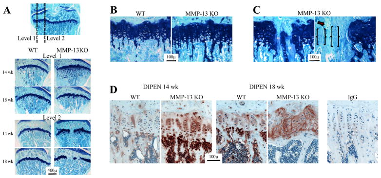
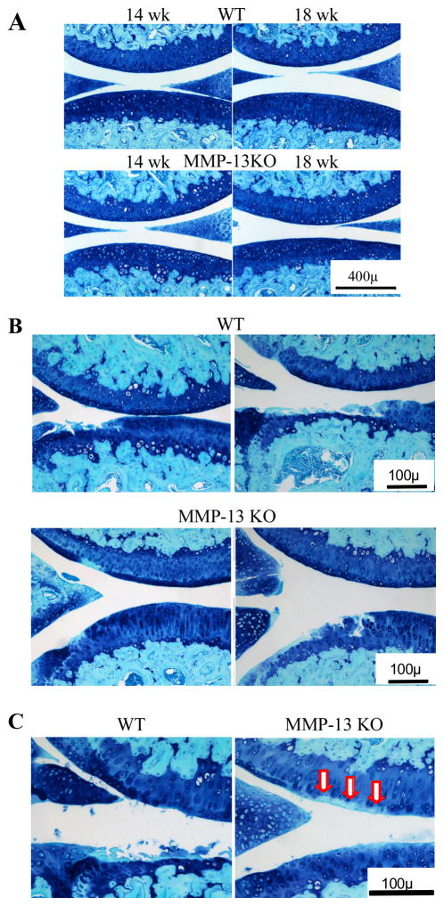
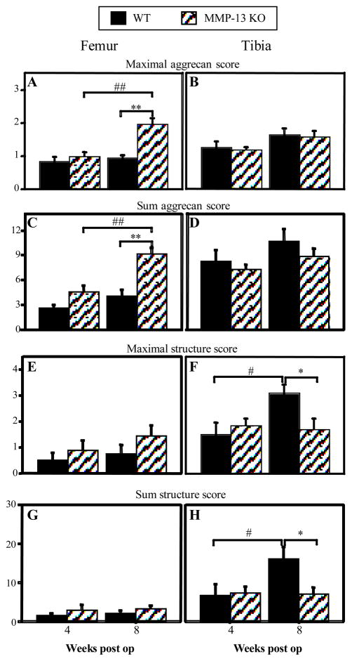
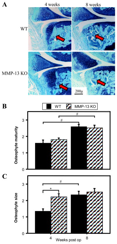
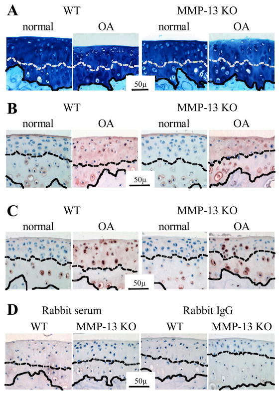
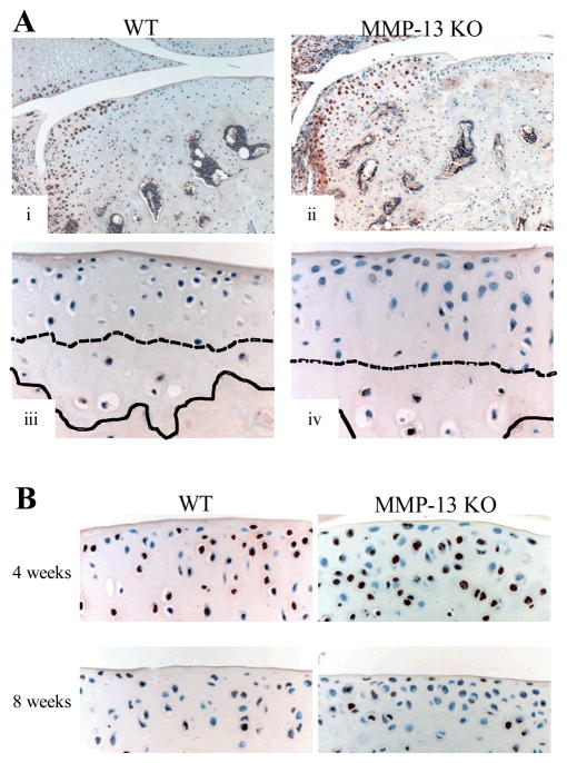
Methods: OA was surgically induced in the knees of MMP-13-knockout mice and wild-type mice, and mice were compared. Histologic scoring of femoral and tibial cartilage aggrecan loss (0-3 scale), erosion (0-7 scale), and chondrocyte hypertrophy (0-1 scale), as well as osteophyte size (0-3 scale) and maturity (0-3 scale) was performed. Serial sections were stained for type X collagen and the MMP-generated aggrecan neoepitope DIPEN.

Results: Following surgery, aggrecan loss and cartilage erosion were more severe in the tibia than femur (P<0.01) and tibial cartilage erosion increased with time (P<0.05) in wild-type mice. Cartilaginous osteophytes were present at 4 weeks and underwent ossification, with size and maturity increasing by 8 weeks (P<0.01). There was no difference between genotypes in aggrecan loss or cartilage erosion at 4 weeks. There was less tibial cartilage erosion in knockout mice than in wild-type mice at 8 weeks (P<0.02). Cartilaginous osteophytes were larger in knockout mice at 4 weeks (P<0.01), but by 8 weeks osteophyte maturity and size were no different from those in wild-type mice. Articular chondrocyte hypertrophy with positive type X collagen and DIPEN staining occurred in both wild-type and knockout mouse joints.

Conclusion: Our findings indicate that structural cartilage damage in a mouse model of OA is dependent on MMP-13 activity. Chondrocyte hypertrophy is not regulated by MMP-13 activity in this model and does not in itself lead to cartilage erosion. MMP-13 deficiency can inhibit cartilage erosion in the presence of aggrecan depletion, supporting the potential for therapeutic intervention in established OA with MMP-13 inhibitors.


Figures

Figure 1
Figure 1
(A) Toluidine blue/fast green stained sections from wild type (WT) and MMP-13 knock out (KO) mice at 14 and 18 weeks of age. The frontal section (top) shows the axial levels from which sagittal sections were collected. Higher magnification images of the growth plate from level 1 (B) shows increased hypertrophic chondrocytes and retention of calcified cartilage trabeculae in the metaphysis of MMP-13 KO mice. In the axial area of MMP-13 KO mice (C) focal areas of growth plate closure had acellular regions with diminished toluidine blue staining (*), faintly stained areas with residual round chondroid-like cells (brackets), and cartilage with no toluidine blue and empty lacunae (arrow). (D) MMP-cleaved aggrecan (DIPEN), was localised to the lower hypertrophic chondrocytes in 14 week-old WT growth plates, but was throughout the growth plate of MMP-13 KO mice at the same age. There was increased DIPEN staining in both genotypes at 18 weeks of age, but it was still more intense in MMP-13 KO compared with WT animals. Minor non-specific staining was observed in sections localized with the same concentration of rabbit IgG as a negative control.
Figure 2
Figure 2
Representative toluidine blue and fast green stained sections from wild type (WT) and MMP-13 knock out (KO) mice at 14 and 18 weeks of age. (A) Central weight-bearing region of the medial femoro-tibial compartment of unoperated joints. (B) Least (left image) and most (right image) severe cartilage erosion in the two genotypes at 8 weeks after surgery are shown. (C) Higher magnification images demonstrating the focal area of aggrecan depletion in the femoral cartilage opposite the tip of the destabilized meniscus in MMP-13 KO (arrows) but not in WT mice, where tibial cartilage erosion is evident.
Figure 3
Figure 3
Maximal (A, B, E, F) and summed (C, D, G, H) scores for aggrecan loss (A–D) and structural cartilage damage (E–H) in femoral and tibial cartilage of wild type (WT) and MMP-13 knock out (KO) mice 4 and 8 weeks after medial meniscal destabilization surgery (“weeks post op”). Graphs represent mean ± SEM; WT n = 12 at 4 and 8 weeks, MMP-13 KO n = 15 at 4 weeks and n = 10 at 8 weeks post op. *p<0.05 **p<0.01 for difference between WT and MMP-13 KO mice at the same time point; #p<0.05 ##p<0.01 for difference between 4 and 8 weeks within a genotype.
Figure 4
Figure 4
(A) Representative toluidine blue & fast green stained sections of wild type (WT) and MMP-13 knock out (KO) mice 4 & 8 weeks after medial meniscal destabilization surgery, demonstrating osteophyte development on the anterior tibial plateau (arrows). The mean scores of osteophyte maturity (B) and osteophyte size (C) in WT and MMP-13 KO mice 4 and 8 weeks after medial meniscal destabilization surgery (“weeks post-op”) are depicted graphically. *p<0.05 for difference between WT and MMP-13 KO mice at the same time point; #p<0.05 for difference between 4 and 8 weeks within a genotype.
Figure 5
Figure 5
Representative sections of proximal tibial cartilage from non-operated (“normal”) joints and after medial meniscal destabilization induced osteoarthritis (“OA”) in wild type (WT) and MMP-13 knock out (KO) mice. (A) Toluidine blue & fast green stain; (B) immunolocalization of type X collagen and (C) immunolocalization of the MMP-generated aggrecan neoepitope DIPEN. Representative negative control sections using equivalent concentration of pre-immune rabbit serum or rabbit IgG in place of the primary antibody in WT and MMP-13 KO mice following surgical induction of OA (D). The dashed line demarcates the tidemark between calcified and non-calcified articular cartilage, while the solid line indicates the osteochondral junction.
Figure 6
Figure 6
(A) Immunolocalization of MMP-14 (brown stain) in the articular cartilage of wild type (WT) and MMP-13 knock out (KO) mice. In both genotypes there was positive immunostaining of chondrocytes in the developing osteophyte (i & iii), and in calcified articular cartilage below the tidemark but not in the non-calcified articular cartilage even in areas with chondrocyte hypertrophy (ii & iv). The dashed line demarcates the tidemark between calcified and non-calcified articular cartilage, while the solid line indicates the osteochondral junction. (B) TUNEL staining (brown) of chondrocytes in the articular cartilage 4 and 8 weeks after surgery in WT and MMP-13 KO mice.

Comment in

References

    1. Pratta M, Yao W, Decicco C, Tortorella M, Liu R, Copeland R, et al. Aggrecan protects cartilage collagen from proteolytic cleavage. J Biol Chem. 2003;278:45539–45545. - PubMed
    1. Caterson B, Flannery CR, Hughes CE, Little CB. Mechanisms involved in cartilage proteoglycan catabolism. Matrix Biol. 2000;19:333–344. - PubMed
    1. Nagase H, Kashiwagi M. Aggrecanases and cartilage matrix degradation. Arthritis Res Ther. 2003;5(2):94–103. - PMC - PubMed
    1. Glasson SS, Askew R, Sheppard B, Carito B, Blanchet T, Ma HL, et al. Deletion of active ADAMTS5 prevents cartilage degradation in a murine model of osteoarthritis. Nature. 2005;434(7033):644–8. - PubMed
    1. Stanton H, Rogerson FM, East CJ, Golub SB, Lawlor KE, Meeker CT, et al. ADAMTS5 is the major aggrecanase in mouse cartilage in vivo and in vitro. Nature. 2005;434(7033):648–52. - PubMed

Publication types

MeSH terms