

Development of a cytotoxic T-cell assay in rabbits to evaluate early immune response to human T-lymphotropic virus type 1 infection
- PMID: 19951176
- PMCID: PMC2852241
- DOI: 10.1089/vim.2009.0059
Development of a cytotoxic T-cell assay in rabbits to evaluate early immune response to human T-lymphotropic virus type 1 infection
Abstract
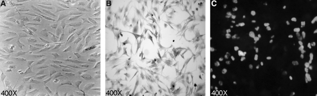
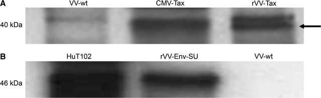
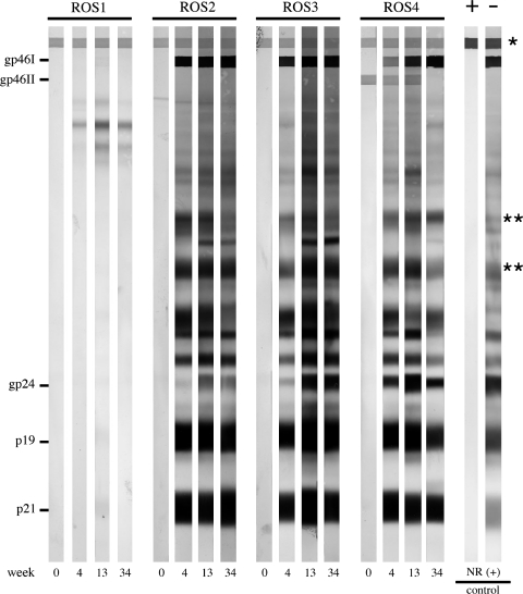
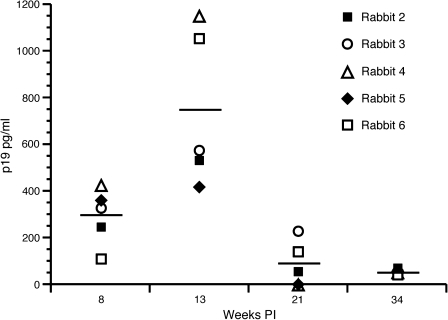
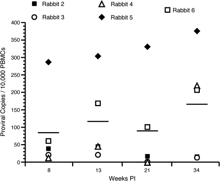
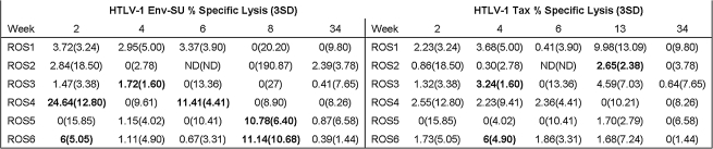
Human T-lymphotropic virus type 1 (HTLV-1) infection causes adult T-cell lymphoma/leukemia (ATL) following a prolonged clinical incubation period, despite a robust adaptive immune response against the virus. Early immune responses that allow establishment of the infection are difficult to study without effective animal models. We have developed a cytotoxic T-lymphocyte (CTL) assay to monitor the early events of HTLV-1 infection in rabbits. Rabbit skin fibroblast cell lines were established by transformation with a plasmid expressing simian virus 40 (SV40) large T antigen and used as autochthonous targets (derived from same individual animal) to measure CTL activity against HTLV-1 infection in rabbits. Recombinant vaccinia virus (rVV) constructs expressing either HTLV-1 envelope surface unit (SU) glycoprotein 46 or Tax proteins were used to infect fibroblast targets in a (51)Cr-release CTL assay. Rabbits inoculated with Jurkat T cells or ACH.2 cells (expressing ACH HTLV-1 molecule clone) were monitored at 0, 2, 4, 6, 8, 13, 21, and 34 wk post-infection. ACH.2-inoculated rabbits were monitored serologically and for viral infected cells following ex vivo culture. Proviral load analysis indicated that rabbits with higher proviral loads had significant CTL activity against HTLV-1 SU as early as 2 wk post-infection, while both low- and high-proviral-load groups had minimal Tax-specific CTL activity throughout the study. This first development of a stringent assay to measure HTLV-1 SU and Tax-specific CTL assay in the rabbit model will enhance immunopathogenesis studies of HTLV-1 infection. Our data suggest that during the early weeks following infection, HTLV-1-specific CTL responses are primarily targeted against Env-SU.
Figures
Similar articles
-
In vivo analysis of replication and immunogenicity of proviral clones of human T-lymphotropic virus type 1 with selective envelope surface-unit mutations.Blood. 2005 Nov 15;106(10):3602-8. doi: 10.1182/blood-2005-03-1076. Epub 2005 Jul 26. Blood. 2005. PMID: 16046523 Free PMC article.
-
Antibodies to the envelope glycoprotein of human T cell leukemia virus type 1 robustly activate cell-mediated cytotoxic responses and directly neutralize viral infectivity at multiple steps of the entry process.J Immunol. 2011 Jul 1;187(1):361-71. doi: 10.4049/jimmunol.1100070. Epub 2011 Jun 6. J Immunol. 2011. PMID: 21646298
-
Functional role of pX open reading frame II of human T-lymphotropic virus type 1 in maintenance of viral loads in vivo.J Virol. 2000 Feb;74(3):1094-100. doi: 10.1128/jvi.74.3.1094-1100.2000. J Virol. 2000. PMID: 10627519 Free PMC article.
-
[Anti-tumor immunity in adult T-cell leukemia].Uirusu. 2004 Jun;54(1):67-74. doi: 10.2222/jsv.54.67. Uirusu. 2004. PMID: 15449906 Review. Japanese.
-
Maintenance of long remission in adult T-cell leukemia by Tax-targeted vaccine: A hope for disease-preventive therapy.Cancer Sci. 2019 Mar;110(3):849-857. doi: 10.1111/cas.13948. Epub 2019 Feb 19. Cancer Sci. 2019. PMID: 30666755 Free PMC article. Review.
Cited by
-
Distinct transformation tropism exhibited by human T lymphotropic virus type 1 (HTLV-1) and HTLV-2 is the result of postinfection T cell clonal expansion.J Virol. 2012 Apr;86(7):3757-66. doi: 10.1128/JVI.06900-11. Epub 2012 Jan 25. J Virol. 2012. PMID: 22278223 Free PMC article.
-
Rabbit Models for Studying Human Infectious Diseases.Comp Med. 2015 Dec;65(6):499-507. Comp Med. 2015. PMID: 26678367 Free PMC article. Review.
-
Human T lymphotropic virus type 1 SU residue 195 plays a role in determining the preferential CD4+ T cell immortalization/transformation tropism.J Virol. 2013 Aug;87(16):9344-52. doi: 10.1128/JVI.01079-13. Epub 2013 Jun 19. J Virol. 2013. PMID: 23785214 Free PMC article.
References
-
- Koksoy S. Phipps AJ. Hayes KA. Mathes LE. SV40 immortalization of feline fibroblasts as targets for MHC-restricted cytotoxic T-cell assays. Vet Immunol Immunopathol. 2001;7:285–295. - PubMed
-
- Lairmore M. Franchini G. Human T-cell leukemia virus types 1, 2. In: Knipe DM, editor. Fields Virology. 5th ed. Wolters Kluwer/Lippincott Williams & Wilkins; Philadelphia, PA: 2007. pp. 2071–2105.
-
- Bertola F. Manigand C. Picard P. Goetz M. Schmitter JM. Precigoux G. N-terminal domain of HTLV-I integrase. Complexation and conformational studies of the zinc finger. J Pept Sci. 20017:588–597. - PubMed
Publication types
MeSH terms
Substances
Grants and funding
LinkOut - more resources
Full Text Sources
