

Hippocampal N-methyl-D-aspartate receptor subunit expression profiles in a mouse model of prenatal alcohol exposure
- PMID: 19951292
- PMCID: PMC3600588
- DOI: 10.1111/j.1530-0277.2009.01096.x
Hippocampal N-methyl-D-aspartate receptor subunit expression profiles in a mouse model of prenatal alcohol exposure
Abstract
Background: Although several reports have been published showing prenatal ethanol exposure is associated with alterations in N-methyl-D-aspartate (NMDA) receptor subunit levels and, in a few cases, subcellular distribution, results of these studies are conflicting.
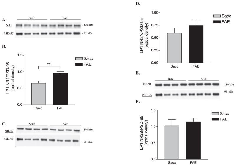
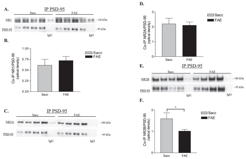
Methods: We used semi-quantitative immunoblotting techniques to analyze NMDA receptor NR1, NR2A, and NR2B subunit levels in the adult mouse hippocampal formation isolated from offspring of dams who consumed moderate amounts of ethanol throughout pregnancy. We employed subcellular fractionation and immunoprecipitation techniques to isolate synaptosomal membrane- and postsynaptic density protein-95 (PSD-95)-associated pools of receptor subunits.
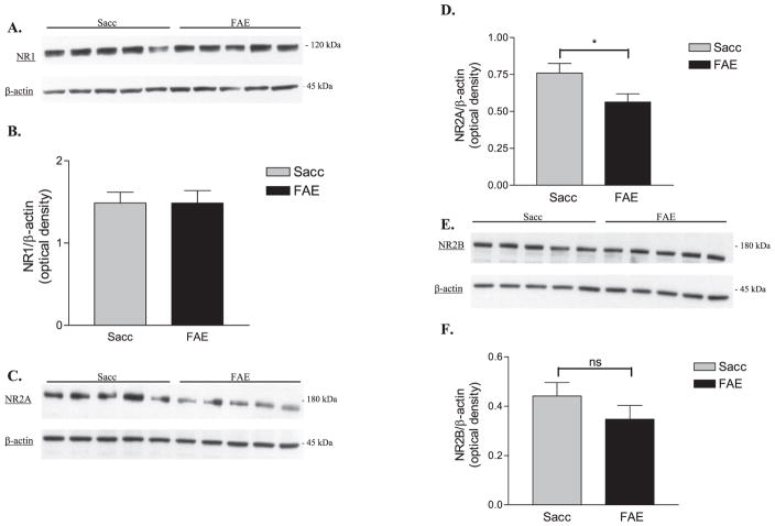
Results: We found that, compared to control animals, fetal alcohol-exposed (FAE) adult mice had: (i) increased synaptosomal membrane NR1 levels with no change in association of this subunit with PSD-95 and no difference in total NR1 expression in tissue homogenates; (ii) decreased NR2A subunit levels in hippocampal homogenates, but no alterations in synaptosomal membrane NR2A levels and no change in NR2A-PSD-95 association; and (iii) no change in tissue homogenate or synaptosomal membrane NR2B levels but a reduction in PSD-95-associated NR2B subunits. No alterations were found in mRNA levels of NMDA receptor subunits suggesting that prenatal alcohol-associated differences in subunit protein levels are the result of differences in post-transcriptional regulation of subunit localization.
Conclusions: Our results demonstrate that prenatal alcohol exposure induces selective changes in NMDA receptor subunit levels in specific subcellular locations in the adult mouse hippocampal formation. Of particular interest is the finding of decreased PSD-95-associated NR2B levels, suggesting that synaptic NR2B-containing NMDA receptor concentrations are reduced in FAE animals. This result is consistent with various biochemical, physiological, and behavioral findings that have been linked with prenatal alcohol exposure.
Figures
Similar articles
-
alpha-Isoform of calcium-calmodulin-dependent protein kinase II and postsynaptic density protein 95 differentially regulate synaptic expression of NR2A- and NR2B-containing N-methyl-d-aspartate receptors in hippocampus.Neuroscience. 2008 Jan 2;151(1):43-55. doi: 10.1016/j.neuroscience.2007.09.075. Epub 2007 Oct 12. Neuroscience. 2008. PMID: 18082335 Free PMC article.
-
Prenatal morphine alters the synaptic complex of postsynaptic density 95 with N-methyl-D-aspartate receptor subunit in hippocampal CA1 subregion of rat offspring leading to long-term cognitive deficits.Neuroscience. 2009 Feb 18;158(4):1326-37. doi: 10.1016/j.neuroscience.2008.11.007. Epub 2008 Nov 8. Neuroscience. 2009. PMID: 19041927
-
Differential interaction of NMDA receptor subtypes with the post-synaptic density-95 family of membrane associated guanylate kinase proteins.J Neurochem. 2008 Feb;104(4):903-13. doi: 10.1111/j.1471-4159.2007.05067.x. J Neurochem. 2008. PMID: 18233995
-
The NR2B subtype of NMDA receptor: a potential target for the treatment of alcohol dependence.Curr Drug Targets CNS Neurol Disord. 2004 Jun;3(3):169-79. doi: 10.2174/1568007043337409. Curr Drug Targets CNS Neurol Disord. 2004. PMID: 15180478 Review.
-
Yotiao, a novel protein of neuromuscular junction and brain that interacts with specific splice variants of NMDA receptor subunit NR1.J Neurosci. 1998 Mar 15;18(6):2017-27. doi: 10.1523/JNEUROSCI.18-06-02017.1998. J Neurosci. 1998. PMID: 9482789 Free PMC article. Review.
Cited by
-
Sex-specific effect of prenatal alcohol exposure on N-methyl-D-aspartate receptor function in orbitofrontal cortex pyramidal neurons of mice.Alcohol Clin Exp Res. 2021 Oct;45(10):1994-2005. doi: 10.1111/acer.14697. Epub 2021 Sep 29. Alcohol Clin Exp Res. 2021. PMID: 34523139 Free PMC article.
-
Long Term Depression in Rat Hippocampus and the Effect of Ethanol during Fetal Life.Brain Sci. 2017 Nov 28;7(12):157. doi: 10.3390/brainsci7120157. Brain Sci. 2017. PMID: 29182556 Free PMC article. Review.
-
Alteration of Gene Expression, DNA Methylation, and Histone Methylation in Free Radical Scavenging Networks in Adult Mouse Hippocampus following Fetal Alcohol Exposure.PLoS One. 2016 May 2;11(5):e0154836. doi: 10.1371/journal.pone.0154836. eCollection 2016. PLoS One. 2016. PMID: 27136348 Free PMC article.
-
Prenatal alcohol exposure alters synaptic activity of adult hippocampal dentate granule cells under conditions of enriched environment.Hippocampus. 2016 Aug;26(8):1078-87. doi: 10.1002/hipo.22588. Epub 2016 Apr 7. Hippocampus. 2016. PMID: 27009742 Free PMC article.
-
Functional and Structural Correlates of Impaired Enrichment-Mediated Adult Hippocampal Neurogenesis in a Mouse Model of Prenatal Alcohol Exposure.Brain Plast. 2020 Dec 29;6(1):67-82. doi: 10.3233/BPL-200112. Brain Plast. 2020. PMID: 33680847 Free PMC article.
References
-
- Abdollah S, Brien JF. Effect of chronic maternal ethanol administration on glutamate and N-methyl-D-aspartate binding sites in the hippocampus of the near-term fetal guinea pig. Alcohol. 1995;12:377–382. - PubMed
-
- Abulrob A, Tauskela JS, Mealing G, Brunette E, Faid K, Stanimirovic D. Protection by cholesterol-extracting cyclodextrins: a role for N-methyl-D-aspartate receptor redistribution. J Neurochem. 2005;92:1477–1486. - PubMed
-
- Al-Hallaq RA, Jarabek BR, Fu Z, Vicini S, Wolfe BB, Yasuda RP. Association of NR3A with N-methyl-D-aspartate receptor NR1 and NR2 subunits. Mol Pharm. 2002;62:1119–1127. - PubMed
-
- Allan AM, Chynoweth J, Tyler LA, Caldwell KK. A mouse model of prenatal ethanol exposure using a voluntary drinking paradigm. Alcohol Clin Exp Res. 2003;27:2009–2016. - PubMed
Publication types
MeSH terms
Substances
Grants and funding
LinkOut - more resources
Full Text Sources
Medical
