. 2009 Dec 4;5(6):610-23.
doi: 10.1016/j.stem.2009.10.019.

SKPs derive from hair follicle precursors and exhibit properties of adult dermal stem cells

Affiliations

SKPs derive from hair follicle precursors and exhibit properties of adult dermal stem cells

Jeffrey Biernaskie et al. Cell Stem Cell. .

Abstract

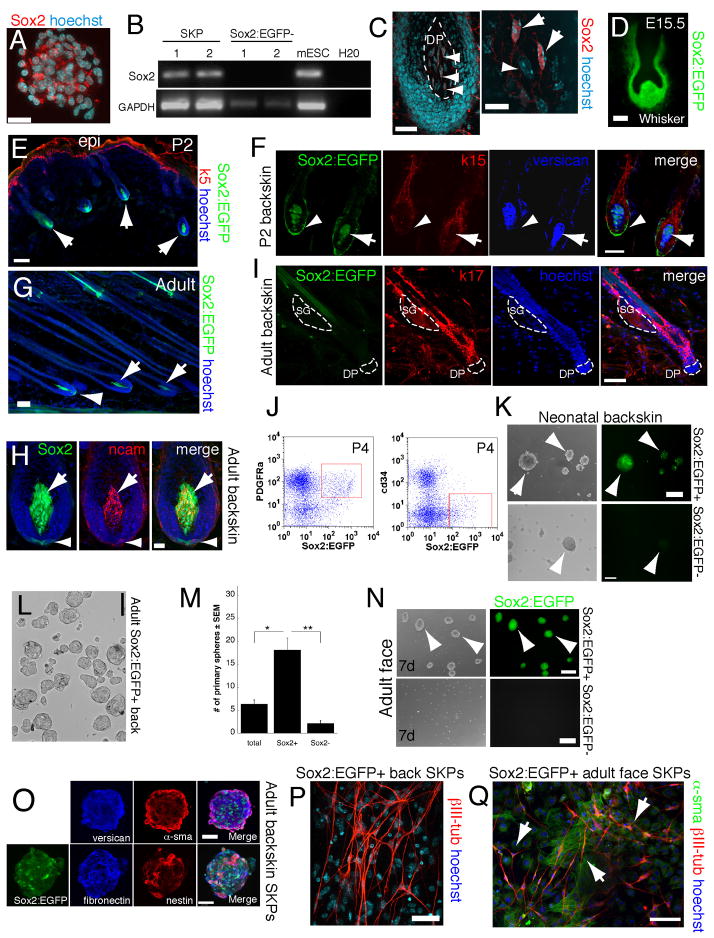
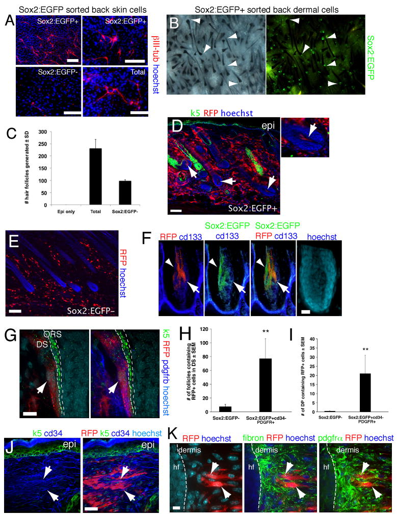
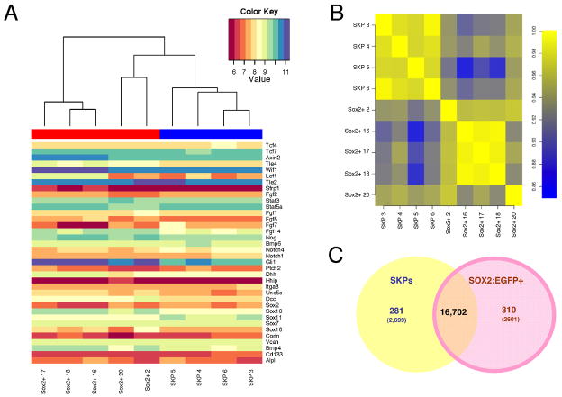
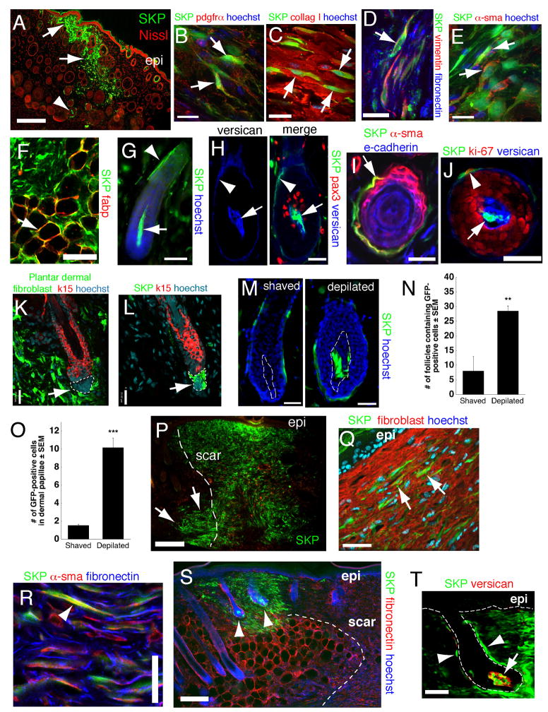
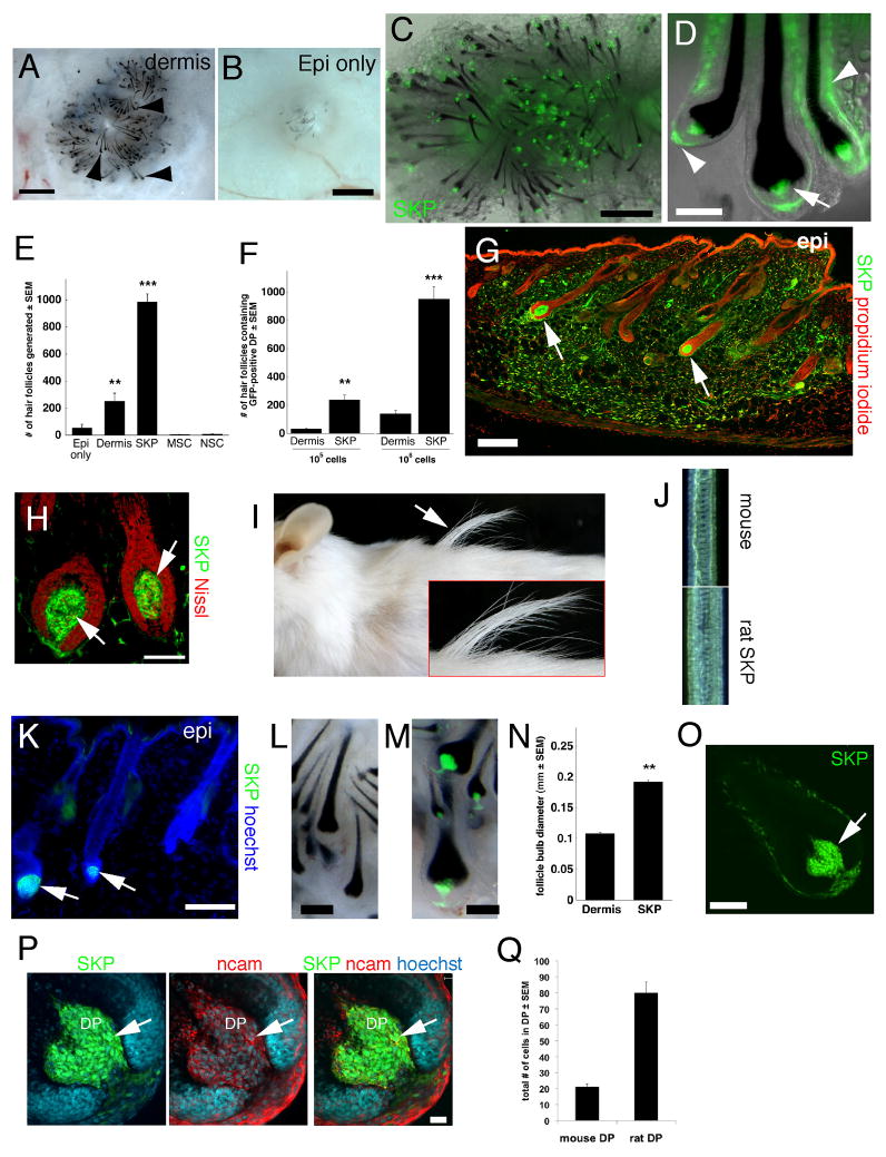
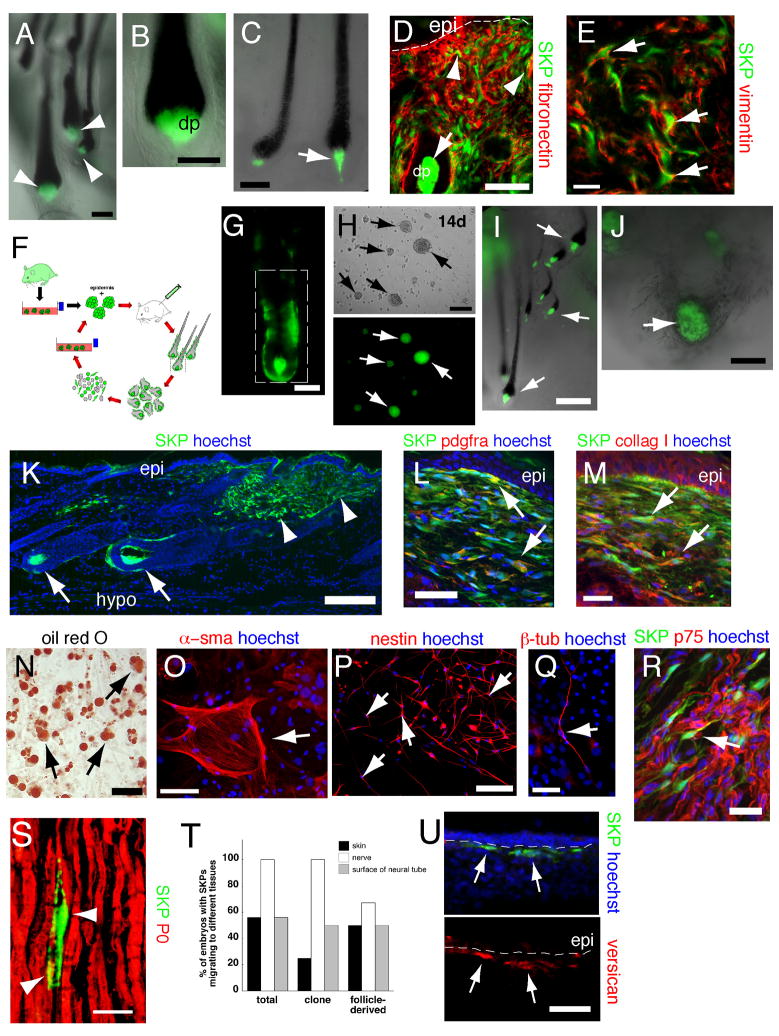
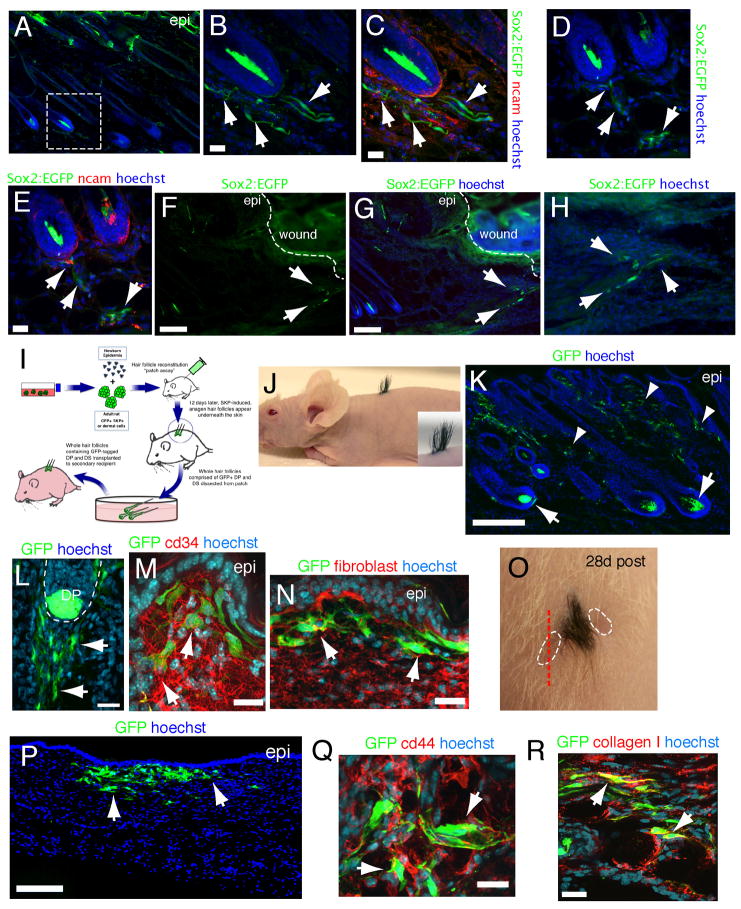
Despite the remarkable regenerative capacity of mammalian skin, an adult dermal stem cell has not yet been identified. Here, we investigated whether skin-derived precursors (SKPs) might fulfill such a role. We show that SKPs derive from Sox2(+) hair follicle dermal cells and that these two cell populations are similar with regard to their transcriptome and functional properties. Both clonal SKPs and endogenous Sox2(+) cells induce hair morphogenesis, differentiate into dermal cell types, and home to a hair follicle niche upon transplantation. Moreover, hair follicle-derived SKPs self-renew, maintain their multipotency, and serially reconstitute hair follicles. Finally, grafting experiments show that follicle-associated dermal cells move out of their niche to contribute cells for dermal maintenance and wound-healing. Thus, SKPs derive from Sox2(+) follicle-associated dermal precursors and display functional properties predicted of a dermal stem cell, contributing to dermal maintenance, wound-healing, and hair follicle morphogenesis.


Figures

Figure 1
Figure 1. Sox2 is dynamically expressed in the hair follicle DP and DS
(A) Neonatal murine SKP sphere immunostained for Sox2 (red). (B) RT-PCR showing Sox2 expression in 2 neonatal murine SKP cultures. Embryonic stem cells (mESC) were a positive control. (C) Longitudinal section of a neonatal mouse follicle showing Sox2+ DP cells (red, arrowheads). Right panel shows Sox2+ (arrows) and negative (arrowhead) DP cells at higher magnification. (D) Sox2:EGFP+ cells (green) in an E15.5 whisker follicle. (E,F) Dorsal skin sections from P2 Sox2:EGFP mice immunostained for EGFP (green), the epidermal markers keratin 5 (E) or keratin 15 (F; both red), and the DP marker versican (blue). In (E) arrows denote anagen hair follicles. In (F) arrowheads indicate EGFP+ DS cells and arrows EGFP+ DP cells. (G–I) Dorsal skin sections of adult Sox2:EGFP mice immunostained for EGFP (green) and NCAM (H, red) or keratin 17 (I, red). (G,H) show EGFP+ cells in anagen hair follicles, with arrows denoting the DP and arrowheads the lower DS. (I) shows a telogen hair follicle where the DP (indicated by hatched lines) is negative for Sox2:EGFP expression. (JQ) Sox2:EGFP+ hair follicle cells proliferate in FGF2 and EGF to generate multipotent SKP spheres. (J) Flow cytometric analysis of neonatal Sox2:EGFP backskin cells sorted for EGFP (x-axis) and PDGFRα or cd34 (y-axis). Red boxes indicate gates for prospective isolation experiments. (K) Sox2:EGFP+ (top panels) and negative (bottom) neonatal backskin cells cultured for 7 days in SKPs conditions. Arrows denote SKP spheres. (L,M) Quantification (M) of SKP spheres generated from sorted adult Sox2:EGFP+ (L) versus Sox2:EGFP− back skin cells in experiments similar to that in (K). Total back skin cells from the same mice were used for comparison (*p<0.05, **p<0.001; n=4). (N) Sox2:EGFP+ (top) and negative (bottom) cells sorted from adult facial skin and cultured in SKPs conditions for 7 days. (O) SKP spheres generated from Sox2:EGFP+ adult back skin cells immunostained for EGFP (green), and the SKP markers fibronectin (blue) and nestin (red; merge is on right) or versican (blue) and α-sma (red; merge is on the right). (P,Q) Sorted adult Sox2:EGFP+ back skin (P) or facial skin (Q) cells were cultured as SKPs, differentiated under neural conditions for 12–18 days and immunostained for the neuronal marker III-tubulin (red in both) and -sma (green in Q). In some panels, cells were counterstained with Hoechst 33258 (blue), as indicated. Scale bars = 25μm in A,C (left panel), 10μm in C (right panel), 50μm in E,F,G,H and 100μm in D,I,K,L,N,O,P,Q. See also Figure S1.
Figure 2
Figure 2. Sox2:EGFP prospectively identifies endogenous dermal precursors that induce hair follicles, home to a hair follicle niche, and differentiate into neural and dermal cell types
(A) Sorted Sox2:EGFP+ and negative neonatal back skin cells differentiated for 12 days under neural conditions, and immunostained for III-tubulin (red). Total cells were sorted but gated only for live cells. (B) Patch assays using sorted Sox2:EGFP+, cd34− neonatal back skin dermal cells. The left panel shows brightfield and the right fluorescence illumination. Arrowheads denote EGFP+ DP. (C) Number of hair follicles generated in patch assays with total or Sox2:EGFP− neonatal dermal cells (n = 3). (D–K) Sorted, uncultured Sox2:EGFP+, cd34−, PDGFRα+ (D,F–K), or Sox2:EGFP− (E) neonatal back skin cells were transplanted into adult NOD/SCID mouse back skin, and analyzed after 2 weeks. All sorted cells were infected with an RFP-expressing retrovirus. (D) Immunostaining for RFP (red) and keratin 5 (green). Arrows denote follicle DP containing transplanted cells, with the right panel at higher magnification. (E) Immunostaining for RFP (red) on skin transplanted with Sox2:EGFP− cells. (F,G) High magnification confocal images of hair follicles immunostained for RFP (F,G, red) and Sox2:EGFP (F, green) plus the DP marker cd133 (F, blue), or keratin-5 (G, green) plus PDGFRβ (G, blue). In (F) the arrowhead and arrow denote Sox2:EGFP+ and cd133+ cells, respectively. In (G) the arrow denotes RFP+, PDGFRβ+ cells in the DS. ORS = outer root sheath. (H,I) Number of hair follicles (H) or follicle DP (I) containing RFP+ transplanted cells as shown in D–G. **P<0.01. (J,K) Immunostaining of interfollicular dermis for RFP (red) and cd34 (J, blue) plus keratin 5 (J, green) or fibronectin (K, pseudocolored green, center panel) plus PDGFRα (pseudocolored green, right panel). Arrows show cells positive for RFP and cd34 (J), or for RFP, fibronectin and PDGFRα (K). hf = hair follicle. Nuclei were stained with Hoechst 33258 (blue), as indicated. Scale bars = 100μm in A,D,E, 25μm in F,J and 10μm in G,K. See also Figure S2.
Figure 3
Figure 3. Microarray analysis demonstrates that SKPs and Sox2:EGFP+ cells are transcriptionally similar
Microarray gene expression profiling of sorted Sox2:EGFP+, cd34− neonatal dermal cells versus primary passage neonatal murine SKPs. (A) Heat map showing the relative expression levels of 36 hair follicle dermal genes in 5 independent Sox2:EGFP cell isolates (red bar on the top left) and 4 independent SKP cultures (blue bar on the top right). Relative expression levels are color-coded as per the color key. The cluster analysis on top shows that some Sox2:EGFP+ cell samples are more related to SKPs than they are to each other. (B) Spearman rank correlations were computed for every pair of microarray experiments. The resulting color-coded correlation matrix reveals the similarity of the transcriptomes of SKPs and Sox2:EGFP+ cells. (C) Venn diagram illustrating that 281/22,102 and 310/22,102 genes are upregulated by at least 2-fold in SKPs and Sox2:EGFP cells respectively, while an additional 2418 and 2291 genes are significantly upregulated by less than 2-fold. See also Figure S3.
Figure 4
Figure 4. SKPs regenerate the dermis and integrate into a hair follicle niche upon transplantation into adult skin
(A,B) Back skin transplanted with YFP-tagged neonatal mouse SKPs 2 weeks earlier and immunostained for YFP (green) and PDGFRα (B, red). Arrows and arrowheads in (A) show transplanted cells in the interfollicular dermis and the DP and DS of follicles, respectively. Arrows in (B) denote double-labeled cells. (C–E) GFP-expressing adult rat SKPs transplanted into depilated adult NOD/SCID mouse dermis 21 days earlier, and immunostained for GFP (green) and collagen type 1 (C, red), vimentin and fibronectin (D, red and blue, respectively), or α-sma (E, red). Arrows denote double-labeled (C,E) or triple-labeled (D) cells. (F) GFP-tagged cells (green) within the hypodermis expressing the adipocyte marker fatty acid binding protein (red, arrow). (G–J) Hair follicles containing neonatal murine YFP+ SKPs 2–4 weeks post-transplantation as in (A). (G) Hair follicle with YFP-labeled cells (green) in the DP (arrow) and DS (arrowhead). (H) Follicle triple-labeled for YFP (green), the DP marker versican (blue) and the melanoblast marker pax3 (red). The arrow and arrowhead indicate the DP and DS. (I,J) Cross-sections of follicles showing transplanted cells (green) in the DS (arrow in I, arrowhead in J) expressing -sma (I, red) or Ki67 (J, red), but not e-cadherin (I, blue, an epidermal marker). In (J), DP cells (arrow) are positive for versican (blue). (K,L) Telogen follicles in skin transplanted 4 weeks earlier with GFP-tagged rat plantar dermal fibroblasts (K) or SKPs (L), immunostained for GFP (green) and keratin 15 (red). Only SKPs are in the DP (arrows, denoted by hatched lines). (M) Representative hair follicles containing neonatal murine YFP+ SKPs (green) 3 weeks after skin was shaved or depilated. Hatched lines denote the DP. (N,O) Number of follicles containing GFP+ cells (N) or GFP+ cells within the DP of individual follicles (O) following transplantation of adult rat GFP+ SKPs into depilated (n=4) versus shaved (n=4) skin. **p<0.01, ***p<0.001. (P–T) Skin 3 (P–R) or 4 (S,T) weeks after transplantation of neonatal YFP-tagged murine SKPs (green in all panels) adjacent to a back skin punch wound. In (P), transplanted cells are present at the site of injection (arrows), and within the regenerated tissue (denoted by dashed lines). (Q,R) Transplanted cells immunostained for fibroblast-specific antigen (Q, red, arrows), or for α-sma and fibronectin (R, arrowhead, red and blue, respectively). (S,T) Transplanted cells are also present in peg-like hair follicles at the boundary of the wound (S, arrowheads) within the DP and DS (T, arrow and arrowheads). Cells in the DP express versican (T, red). Some sections were counterstained with Hoechst 33258 (blue) or fluorescent Nissl (red), as indicated. Epi = epidermis. Scale bars = 200μm in A,P,S, 16μm in B–E, 25 μm in G-M,Q, 50μm in F,R,T. See also Figure S4.
Figure 5
Figure 5. SKPs, but not other adult stem cells, reconstitute a hair follicle niche and instruct epidermal cells to generate hair follicles
(A,B), Patch assays at 12 days, combining neonatal C57/Bl6 murine epidermal cells with (A) and without (B) 106 neonatal rat dermal cells, visualized by phase illumination (arrowheads denote hair follicles, which are black). (C,D) Combined phase and fluorescence illumination of similar patch assays with 106 GFP-expressing adult rat SKPs (green) (arrow and arrowheads in D denote the DP and DS, respectively). (E) Number of hair follicles with GFP+ or YFP+ DP as shown in (C,D), using 106 adult rat GFP+ SKPs or MSCs, or neonatal murine YFP+ dermal cells or NSCs. ** p<0.001 relative to epidermis only, ***p<0.001 relative to epidermis only and to dermis (n=6 for adult rat SKPs, and 3 for other groups). (F) Quantification of hair follicle numbers as in (E), where adult rat SKPs were compared to neonatal rat dermal cells. ***p<0.001 relative to dermal cells (n=3 and 4 each for 105 and 106 cells, respectively). (G–K) Adult GFP-expressing rat SKPs (green) were injected into the back skin of adult NOD/SCID mice for 8 weeks. (G,H) Immunostaining for GFP (green). Arrows indicate transplanted cells in hair follicle anagen DP. (I,J) Chimeric rat/mouse hairs were longer (I) and thicker (J) than endogenous pelage hairs. (K) Some transplanted GFP+ cells comprised the DP of telogen hair follicles (arrows). (L,M) Hair follicles from patch assays where 106 mouse neonatal dermal cells (L) or dissociated adult rat GFP-expressing SKPs (M, green) were used. (N) Hair bulb diameter in follicles similar to those in (L,M) (n=2 independent experiments). **p=0.0074. (O–Q) Hair follicles were generated in patch assays using either YFP+ mouse or GFP+ rat SKPs. (O,P) A single hair follicle derived from rat SKPs at low (O) and high (P) magnification, immunostained for GFP (green), NCAM (red), which marks DP and DS cells, and Hoechst to delineate cell nuclei. (Q) Number of cells in the DP of follicles generated from mouse versus rat SKPs, as shown in (P). Nuclei were stained with propidium iodide (red), fluorescent Nissl (red) or Hoechst 33258 (blue), as indicated. epi=epidermis, hypo=hypodermis. Scale bars = 500μm in A,C, 1mm in B, 50μm in D,H, 100μm in G,K,L,M,O and 20μm in P. See also Figure S5.
Figure 6
Figure 6. (A–E) Clonally-derived SKPs reconstitute the dermis and induce hair follicle formation
Analysis of one adult rat GFP+ SKP clone in patch assays (A–C), or by transplantation into adult mouse dermis (D,E). After 2–4 (A,B) or 11 months (C) of culturing, clonal SKPs (green) induced the formation of hair follicles, where they comprised the DS and DP (arrowheads in A, arrow in C). (D,E) After 12 weeks in culture, cells from the same clone were transplanted and skin immunostained for GFP (green) and fibronectin (D, red, arrowheads) or vimentin (E, red, arrows). Arrow in (D) denotes the DP. (FU) SKPs isolated from their hair follicle niche self-renew, serially reconstitute hair follicles and remain multipotent. (F) Schematic showing the serial reconstitution assay of hair morphogenesis. (G,H) A follicle isolated from a patch assay using adult rat GFP-labeled cells. Tagged cells isolated from the boxed area generated GFP+ SKP spheres (H, arrows) after 12 days in culture as seen by phase (top) and fluorescence (bottom) illumination. (I) Cells from these follicle-derived spheres induced formation of secondary hair follicles in the patch assay (arrows). (J) In tertiary follicle reconstitutions, follicle-derived SKPs generated hair follicles, but many also aggregated into DP-like structures surrounded by black melanocytes (arrow). (K–M) Skin transplanted with GFP+ follicle-derived SKPs for 4 weeks. Transplanted cells (green) integrated into follicle DS and DP (K, arrows) and contributed to the dermis (K, arrowheads), where they expressed PDGFRα (L, red) and collagen type 1 (M, red). Arrows indicate double-labeled cells. (N,O) Follicle-derived SKPs generated adipocytes, as indicated by the lipophilic dye oil red O (N, arrows), and sma+ cells, potentially myofibroblasts (O, arrow) in mesodermal differentiations. (P,Q) When differentiated under neurogenic conditions, they generated nestin+ cells after 5 days (P, red, arrows), and III-tubulin positive cells after 14 days (Q, red, arrow). (R,S) Sciatic nerve sections 6 weeks after transplant of follicle-derived SKPs, showing that some transplanted cells expressed the Schwann cell markers p75NTR (R, arrow) and P0 (S, arrowheads). (T,U) Transplantation of follicle-derived SKPs into the chick neural crest migratory stream (H.H. stage 18). (T) Quantification after 3 days in ovo showed follicle-derived (n=6) and clonal (n=8) SKPs behaved like total SKPs (n=9), migrating to the ventral nerve or DRG and skin, with some remaining close to the neural tube. (U) After 8 days in ovo, some of the transplanted cells that were in the dermis (green) were versican+ (red, arrows). Nuclei were stained with Hoechst 33258 (blue), as indicated. epi = epidermis. Scale bars = 100μm in A,B,C,D,H,P, 50μm in E,G,N,O,Q,U, 25μm in L,M,R,S, 200um in I,K, and 250μm in J.
Figure 7
Figure 7. (A–H) Sox2:EGFP+ cells migrate out of hair follicles following punch wounds
Immunostaining for EGFP (green) and NCAM (C,E, red) in Sox2:EGFP skin 3 days after a punch wound. (B–E) show hair follicles in the vicinity of the injury, with (B,C) being higher magnification images of the boxed area in (A), while (D,E) show the same field. (F––H) show the region directly adjacent to the wound, with (H) being a higher magnification image. Arrows show Sox2:EGFP+ cells outside of hair follicles. (IR) Lineage tracing demonstrates that follicular DP and DS cells contribute to dermal maintenance and wound healing. (I) Schematic depicting lineage tracing procedure. Hair follicles were generated in patch assays using GFP-tagged rat SKPs or uncultured neonatal rat dermal cells, and 40–60 newly-formed genetically-tagged follicles were carefully dissected and grafted on to the back skin of a recipient mouse. Skin was analyzed 4–8 weeks later. (J) Photograph of an adult nude mouse that had received a transplant of chimeric hair follicles as in (I) 3 weeks earlier. (K–N) Back skin of a mouse as in (I) 4 weeks post-transplant immunostained for GFP (green) and cd34 (M, red, arrows) or fibroblast-specific antigen (N, red, arrows). Arrows and arrowheads in (K) denote GFP-tagged cells in follicle DP and DS and interfollicular dermis, respectively. Arrows in (L) denote GFP+ cells leaving the hair follicle. (O) Photograph of a transplant similar to (J), except that two adjacent punch wounds (hatched lines) were made 3 weeks after the initial hair follicle graft, and skin was analyzed 4 weeks later. (P–R) Skin sectioned as shown by the red hatched line in (O), and immunostained for GFP (green) and cd44 (red, Q) or collagen type I (red, R). Arrows denote transplanted cells in P and double-labeled cells in Q,R. Nuclei were stained with Hoechst 33258 (blue). Scale bars = 200um in K,P and 25μm in B,C,E,L,M,N,Q,R. See also Figure S7.

Comment in

Similar articles

Cited by

References

    1. Avilion AA, Nicolis SK, Pevny LH, Perez L, Vivian N, Lovell-Badge R. Multipotent cell lineages in early mouse development depend on SOX2 function. Genes Dev. 2003;17:126–140. - PMC - PubMed
    1. Biernaskie J, McKenzie IA, Toma JG, Miller FD. Isolation of skin-derived precursors (SKPs) and differentiation and enrichment of their Schwann cell progeny. Nat Protocols. 2006;1:2803–2812. - PubMed
    1. Driskell RR, Giangreco A, Jensen KB, Mulder KW, Watt FM. Sox2-positive dermal papilla cells specify hair follicle type in mammalian epidermis. Development. 2009;136:2815–2823. - PMC - PubMed
    1. Ellis P, Fagan BM, Magness ST, Hutton S, Taranova O, Hayashi S, McMahon A, Rao M, Pevny L. SOX2, a persistent marker for multipotential neural stem cells derived from embryonic stem cells, the embryo or the adult. Dev Neurosci. 2004;26:148–165. - PubMed
    1. Fernandes KJ, Kobayashi NR, Gallagher CJ, Barnabe-Heider F, Aumont A, Kaplan DR, Miller FD. Analysis of the neurogenic potential of multipotent skin-derived precursors. Exp Neurol. 2006;201:32–48. - PubMed

Publication types

Substances

Associated data