. 2009 Nov 30;187(5):655-68.
doi: 10.1083/jcb.200908036.

Ube2j2 ubiquitinates hydroxylated amino acids on ER-associated degradation substrates

Affiliations

Ube2j2 ubiquitinates hydroxylated amino acids on ER-associated degradation substrates

Xiaoli Wang et al. J Cell Biol. .

Abstract

Ubiquitin (Ub) modification of proteins plays a prominent role in the regulation of multiple cell processes, including endoplasmic reticulum-associated degradation (ERAD). Until recently, ubiquitination of substrates was thought to occur only via isopeptide bonds, typically to lysine residues. Several recent studies suggest that Ub can also be coupled to nonlysine residues by ester/thiolester bonds; however, the molecular basis for these novel modifications remains elusive. To probe the mechanism and importance of nonlysine ubiquitination, we have studied the viral ligase murine K3 (mK3), which facilitates the polyubiquitination of hydroxylated amino acids serine/threonine on its ERAD substrate. In this paper, we identify Ube2j2 as the primary cellular E2 recruited by the mK3 ligase, and this E2-E3 pair is capable of conjugating Ub on lysine or serine residues of substrates. However, surprisingly, Ube2j2-mK3 preferentially promotes ubiquitination of hydroxylated amino acids via ester bonds even when lysine residues are present on wild-type substrates, thus establishing physiological relevance of this novel ubiquitination strategy.


Figures

Figure 1.
Figure 1.
Membrane-bound E2s support the ubiquitination of the HC by mK3. (A) Mouse WT3 cells coexpressing WT Ld and mK3 were treated briefly with digitonin. The cytosolic proteins (CP) in solution and membrane proteins in the pellet were separated by centrifugation. The pellet was then suspended with reaction buffer containing an ATP-regenerating system. Three aliquots were made from this suspension and incubated with E1 only, E1 + HA-Ub (Ub*), or rabbit reticulocyte FII + Ub*, respectively, and followed by centrifugation. Samples from supernatant (S) and pellet (P) of each group as well as the cytosolic protein fraction were blotted by the antibodies indicated to verify the depletion of the cytosolic proteins and the maintenance of membrane proteins in the pellet. It should be noted that FII likely contains most soluble E2s; thus, the supernatant fraction with FII added is Ube2k and Ube2n positive. (B) Precipitations of Ld HCs from the supernatant or the pellet fractions of three reaction groups described in A were performed using anti-Ld mAbs. The precipitates were blotted for newly formed Ub-Ld conjugates with anti-HA. An anti-Ld blot was included to show similar input in each group and as an Endo H treatment control. Data shown are representative of three independent experiments. (A and B) Molecular mass is indicated in kilodaltons. IB, immunoblot; IP, immunoprecipitation.
Figure 2.
Figure 2.
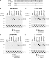
Ube2j2 interacts with mK3 to promote the Ub conjugation of S or K residues, whereas Ube2d1 interacts with mK3 to promote only ubiquitination of K residues. (A) Nomenclature and sequence alignment of the cytoplasmic tails of Ld mutants are shown with original WT residues in bold. (B) WT3 cells coexpressing Ld 329K (1K) and mK3 were permeabilized, cytosol depleted, and incubated with E1 and selective mouse E2 in the presence of ATP and HA-Ub. Precipitated Ld molecules (−/+ Endo H) were resolved by SDS-PAGE and blotted for newly ubiquitinated Ld by anti-HA. An anti-Ld blot was included to show similar input in each group and as an Endo H treatment control. (C) After permeabilization and centrifugation, cells used in B were incubated with one of the four ER-associated E2s or homologues. Precipitates of Ld HCs were blotted for HA-Ub and Ld. (D and E) The same experiments were conducted as in B and C, respectively, using cells stably expressing Ld 329S (1S) and mK3. (B–E) Data are representative of at least two independent experiments. Molecular mass is indicated in kilodaltons. IB, immunoblot; IP, immunoprecipitation.
Figure 3.
Figure 3.
Ube2j2 is required for mK3-induced HC ubiquitination and degradation. (A) WT3 cells expressing WT Ld and mK3 were transduced by lentivirus-based shRNA targeting one of the following Ub-conjugating enzymes: Ube2d1, Ube2g1, Ube2g2, Ube2j1, or Ube2j2 (S#1). After incubation with 50 µM MG132 for 2 h, Ld HCs were precipitated from these cells and followed by SDS-PAGE and blotting with anti-Ub and anti-Ld antibodies. The β-actin and mK3 blots in the middle panel were included to show a similar amount of input lysate and mK3 in each cell line. In the right panel, selected cell lines from the left panel were incubated for 24 h with 125 U/ml IFN-γ, pulse labeled with [35S]Cys/Met, and chased for the indicated times with unlabeled Cys/Met. Ld precipitates were resolved by SDS-PAGE and visualized by autoradiography. Relative band intensities from the gels are plotted as a percentage of the intensity at time 0 for each cell line. Data are representative of three independent experiments. (B) WT3 cells expressing WT Ld and mK3 were transduced by retrovirus-based shRNA targeting Ube2j2 (S#2) or Ube2n or by an empty vector. The ubiquitination and degradation of Ld HCs were determined by immunoblotting and pulse–chase experiments as in A. Data are representative of three independent experiments. (C) Selected cells used in A and B were permeabilized, cytosol depleted, and incubated with E1 or E1 + Ube2j2 in the presence of ATP, HA-Ub, and Ub-Al. Precipitated Ld molecules were resolved by SDS-PAGE and blotted for newly ubiquitinated Ld by anti-HA. An anti-Ld blot was included to show the level of Ld molecules in each cell line. (D) Same as in C except that WT3 cells expressing 1S Ld and mK3 were used. (A–D) Molecular mass is indicated in kilodaltons. IB, immunoblot; IP, immunoprecipitation.
Figure 4.
Figure 4.
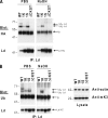
Ub conjugates on WT Ld molecules induced by mK3 are sensitive to mild alkaline treatment. (A) WT3 cells expressing mK3 and WT Ld or one of the mutants Ld 1K, Ld 1S, or Ld ΔCKST were permeabilized, cytosol depleted, and incubated with E1, Ube2j2, and HA-Ub. After treatment with PBS or 0.1 M NaOH for 1 h at 37°C, Ld HC precipitates from these cells were neutralized and resolved by reducing SDS-PAGE and then blotted with anti-HA and anti-Ld. A background Ig heavy chain band can be seen in the blot. Data are representative of two independent experiments. (B) Ld HCs were precipitated from cells used in A without permeabilization. After NaOH treatment, SDS-PAGE and immunoblotting were conducted as in A, except using anti-Ub to visualize ubiquitinated Ld HC. The β-actin and mK3 blots on the right were included to show a similar amount of input lysate and mK3 in each cell line. Data are representative of three independent experiments. (A and B) Molecular mass is indicated in kilodaltons. IP, immunoprecipitation.
Figure 5.
Figure 5.
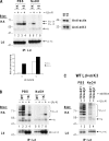
Ube2j2–mK3 favors ubiquitination of S residues over K residues on the tail of HC substrates. (A) WT3 cells expressing mK3 and Ld 1K (329K) or Ld 1S (329S) were permeabilized, cytosol depleted, and incubated with E1, Ube2j2, and HA-Ub in the presence or absence of 2 µM Ub-Al, a DUB inhibitor. After treatment with PBS or NaOH for 45 min, Ld HC precipitates from these cells were neutralized and resolved by reducing SDS-PAGE and then blotted with anti-HA and anti-Ld. Background Ig heavy chain bands can be seen in the blot. Quantification of Ub2-Ld bands from PBS treatment in the gel is shown in the graph. Samples from cytosolic proteins and pellets of each line were blotted to verify an equal amount of mK3 expression (right). (B) WT3 cells expressing mK3 and WT Ld or Ld 1K were permeabilized and incubated with E1, Ube2j2, and HA-Ub in the presence or absence of Ub-Al. All other steps were performed as in A. (C) Same as in A, except permeabilized cells expressing mK3 and WT Ld were incubated with either Ube2d1 or Ube2j2 in the presence of Ub-Al. Data shown are representative of two independent experiments. (A–C) Molecular mass is indicated in kilodaltons. IP, immunoprecipitation.
Figure 6.
Figure 6.
The kK3 and kK5 RINGs in the context of mK3 interact with Ube2j2 to promote the formation of K48-linked Ub chain on S residues of the HCs. (A) kK3 or kK5 RING-mK3 chimeric molecules were expressed in WT3–Ld 1S cells. After permeabilization and depletion of cytosolic proteins, aliquots of these cells were incubated with no enzyme, E1 only, or E1 plus one of the two E2s in the presence of ATP and HA-Ub. Ld HCs were immunoprecipitated and blotted as indicated. (B) Aliquots of the cells used in A were incubated with ATP, E1, and WT or mutant HA-Ub in an ubiquitination assay of permeabilized cells as described in A. Immunoprecipitation (IP) of Ld and blotting by anti-HA (top) or anti-Ub (middle) antibodies shows newly formed Ub-Ld HCs and total Ub-Ld HCs, respectively. The conjugates formed by HA-Ub are in red. (A and B) Molecular mass is indicated in kilodaltons. IB, immunoblot.

Similar articles

Cited by

References

    1. Amerik A.Y., Hochstrasser M. 2004. Mechanism and function of deubiquitinating enzymes. Biochim. Biophys. Acta. 1695:189–207 10.1016/j.bbamcr.2004.10.003 - DOI - PubMed
    1. Arteaga M.F., Wang L., Ravid T., Hochstrasser M., Canessa C.M. 2006. An amphipathic helix targets serum and glucocorticoid-induced kinase 1 to the endoplasmic reticulum-associated ubiquitin-conjugation machinery. Proc. Natl. Acad. Sci. USA. 103:11178–11183 10.1073/pnas.0604816103 - DOI - PMC - PubMed
    1. Bays N.W., Gardner R.G., Seelig L.P., Joazeiro C.A., Hampton R.Y. 2001. Hrd1p/Der3p is a membrane-anchored ubiquitin ligase required for ER-associated degradation. Nat. Cell Biol. 3:24–29 10.1038/35050524 - DOI - PubMed
    1. Biederer T., Volkwein C., Sommer T. 1997. Role of Cue1p in ubiquitination and degradation at the ER surface. Science. 278:1806–1809 10.1126/science.278.5344.1806 - DOI - PubMed
    1. Boname J.M., Stevenson P.G. 2001. MHC class I ubiquitination by a viral PHD/LAP finger protein. Immunity. 15:627–636 10.1016/S1074-7613(01)00213-8 - DOI - PubMed

Publication types

MeSH terms