. 2009 Dec 1;23(23):2729-41.
doi: 10.1101/gad.1824509.

YAP1 is amplified and up-regulated in hedgehog-associated medulloblastomas and mediates Sonic hedgehog-driven neural precursor proliferation

Affiliations

YAP1 is amplified and up-regulated in hedgehog-associated medulloblastomas and mediates Sonic hedgehog-driven neural precursor proliferation

Africa Fernandez-L et al. Genes Dev. .

Abstract

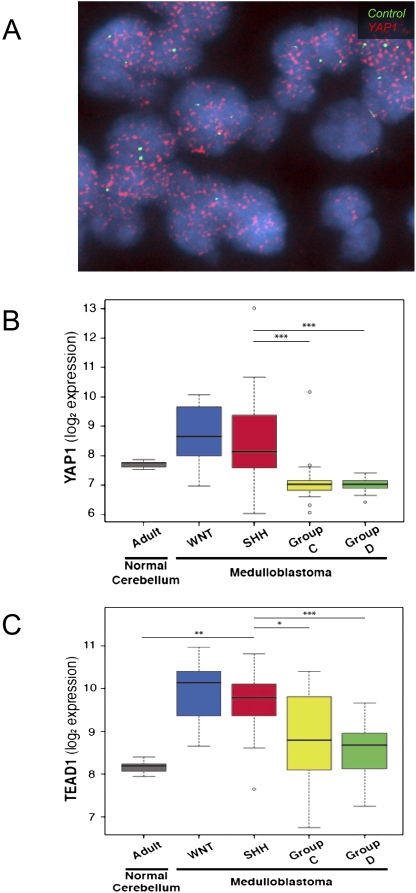
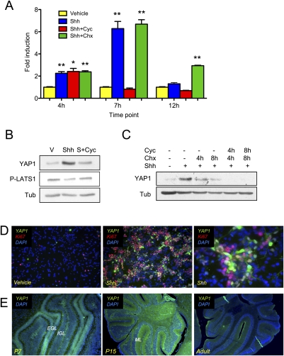
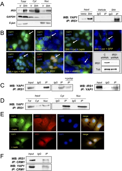
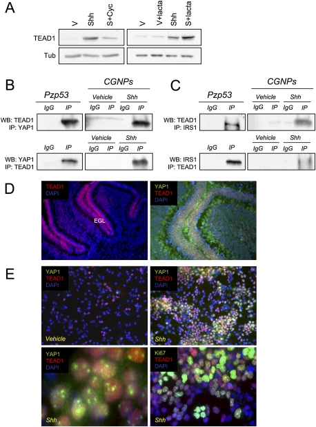
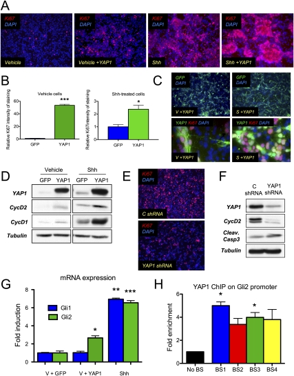
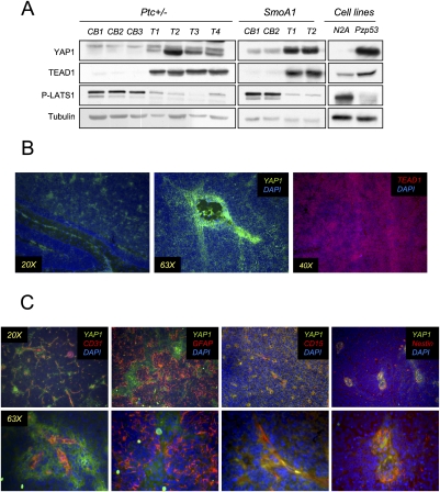
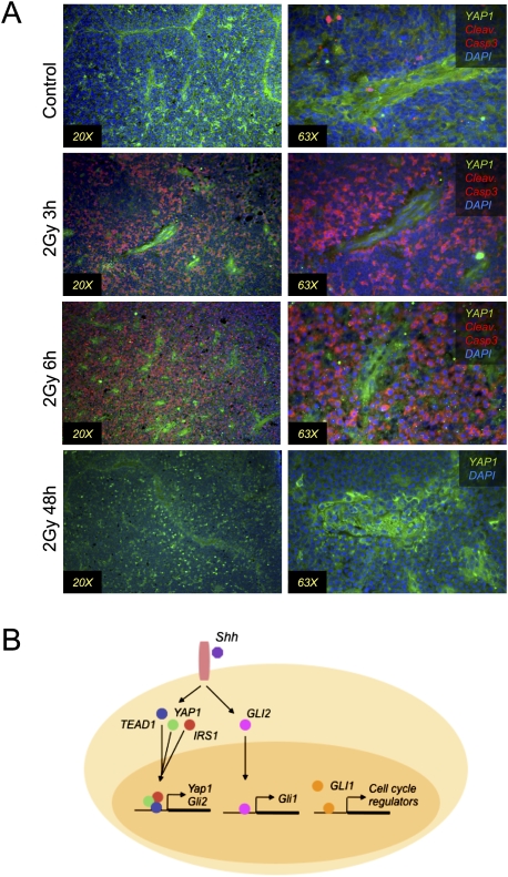
Medulloblastoma is the most common solid malignancy of childhood, with treatment side effects reducing survivors' quality of life and lethality being associated with tumor recurrence. Activation of the Sonic hedgehog (Shh) signaling pathway is implicated in human medulloblastomas. Cerebellar granule neuron precursors (CGNPs) depend on signaling by the morphogen Shh for expansion during development, and have been suggested as a cell of origin for certain medulloblastomas. Mechanisms contributing to Shh pathway-mediated proliferation and transformation remain poorly understood. We investigated interactions between Shh signaling and the recently described tumor-suppressive Hippo pathway in the developing brain and medulloblastomas. We report up-regulation of the oncogenic transcriptional coactivator yes-associated protein 1 (YAP1), which is negatively regulated by the Hippo pathway, in human medulloblastomas with aberrant Shh signaling. Consistent with conserved mechanisms between brain tumorigenesis and development, Shh induces YAP1 expression in CGNPs. Shh also promotes YAP1 nuclear localization in CGNPs, and YAP1 can drive CGNP proliferation. Furthermore, YAP1 is found in cells of the perivascular niche, where proposed tumor-repopulating cells reside. Post-irradiation, YAP1 was found in newly growing tumor cells. These findings implicate YAP1 as a new Shh effector that may be targeted by medulloblastoma therapies aimed at eliminating medulloblastoma recurrence.


Figures

Figure 1.
Figure 1.
YAP1 is amplified in a subset of human medulloblastomas, and YAP1 and TEAD1 are overexpressed in human Shh- and Wnt-driven medulloblastomas. (A) Interphase FISH with a YAP1 probe (red signal) revealed a double-minute pattern of hybridization in two out of 67 (3%) human medulloblastomas. A control probe targeting 11p11.2 (green signal) showed a normal complement. (B) Box plot showing YAP1 mRNA expression obtained from an exon array profiling of 110 human medulloblastomas and 14 cerebella. YAP1 is highly expressed in SHH- and WNT-driven medulloblastomas. These plots are a useful means of displaying differences between populations (i.e., medulloblastoma subgroups) as they depict groups of numerical data (in this case, signal intensity/expression level for the respective genes in adult cerebellum and medulloblastoma subgroups) through their five-number summaries: the smallest observation (sample minimum = lower line), lower quartile (Q1 = bottom of box), median (Q2 = line in box), upper quartile (Q3 = top of box), and largest observation (sample maximum = upper line). These plots are also able to identify any observations that may represent outliers (circles outside the boxes). Note the outlier above the YAP1 SHH plot is a medulloblastoma with genomic amplification of YAP1 and concordant YAP1 expression. (C) Box plot showing TEAD1 mRNA expression in the same sample series. Note high levels of expression in WNT- and SHH-associated medulloblastomas. To statistically compare the expression of YAP1 and TEAD1 in SHH-driven medulloblastomas to their relative expression in the individual subgroups (and to the normal adult cerebellum), we performed the Wilcoxon rank-sum (Mann-Whitney) test. Statistically significant differences are indicated as (*) P < 0.01; (**) P < 0.001; (***) P < 0.0001. These results show that YAP1 and TEAD1 expression is significantly higher in SHH-driven (and WNT-driven) medulloblastoma than Group C and Group D tumors. Similar results of significance were obtained when comparing YAP1 and TEAD1 expression in WNT medulloblastoma to either adult cerebellum or GroupC/D tumors.
Figure 2.
Figure 2.
YAP1 mRNA and protein expression are up-regulated by Shh. (A) YAP1 mRNA expression in CGNPs treated as indicated was analyzed by real-time PCR and fold change is represented. There is a transient increase in YAP1 mRNA expression in Shh-treated cells with a maximum increase at 7 h. Treatment with cyclopamine (cyc) blocks YAP1 mRNA up-regulation, while treatment with cycloheximide (chx) does not. Statistically significant differences are indicated as (*) P < 0.05; (**) P < 0.01; (***) P < 0.001. (B) CGNPs were cultured in vitro for 48 h in the presence or absence of Shh, then treated with cyclopamine for 12 h when indicated. Shh treatment leads to YAP1 protein accumulation and reduced LATS1 phosphorylation. Both events are prevented in the presence of cyclopamine. (C) CGNPs were cultured in vitro for 48 h in the presence or absence of Shh ± cyclopamine and/or cycloheximide. Treatment with cyclopamine accelerates YAP1 protein degradation. (D) CGNPs cultured in the absence or presence of Shh were fixed and immunostained for YAP1 and Ki67. (Left) CGNPs expressing YAP1 are also Ki67 positive. (E) Cerebella of SW129 mice were immunostained for YAP1 at three developmental stages. (Left) At P7, YAP1 is expressed mainly in the EGL. (Middle) At P15, YAP1 is present in granule cells in the IGL, and in their processes in the molecular layer. In adult cerebella, we also detect YAP1 protein distributed sparsely throughout the IGL.
Figure 3.
Figure 3.
IRS1 interacts with YAP1 and regulates its nuclear accumulation. (A) Subcellular fractionation of CGNPs cultured in the presence or absence of Shh. (Left) The greatest accumulation of IRS1 protein after Shh treatment takes place in the nucleus. (Right) Immunoprecipitation of IRS1 in Shh-treated CGNPs brings down YAP1. (B, top panel, left) Immunostaining shows that YAP1 is mainly nuclear in Shh-treated CGNPs. (Top panel, second from left) In the presence of cyclopamine, YAP1 is excluded from the nucleus. (Top panel, second from right) In the presence of cyclopamine and leptomycin, YAP1 accumulates in the nucleus. Overexpressing IRS1 in the presence of cyclopamine leads to YAP1 accumulation in the nucleus (bottom panel, left), while overexpressing control viruses (GFP) does not (top panel, right). (Bottom panel, second from left) Cells that get infected with IRS1 (GFP reporter) viruses have nuclear YAP1. (Bottom panel, second from right) IRS1 knockdown prevents YAP1 from accumulating in the nucleus. (Bottom panel, right) The knockdown efficiency is shown by Western blot. (C) Immunoprecipitation of IRS1 in the Pzp53 medulloblastoma cell line coprecipitates YAP1. (Left) The YAP1:IRS1 interaction was decreased in the presence of cyclopamine. (Right) IRS1 was also detected in YAP1 immunoprecipitates. (D) Subcellular fractionation of Pzp53 cells and subsequent IRS1 immunoprecipitation. YAP1 was detected in both the nuclear and cytoplasmic precipitates. (E) Pzp53med cells were treated with leptomycin, fixed, and immunostained for YAP1 and IRS1. (Top row) In untreated cells, YAP1 and IRS1 are both nuclear and cytoplasmic. (Bottom row) In leptomycin-treated cells, there is an accumulation of both proteins in the nucleus. (F) Immunoprecipitation of CRM1 in Pzp53 cells. Both YAP1 and IRS1 were detected in the immunoprecipitate.
Figure 4.
Figure 4.
TEAD1 interacts with YAP1 in CGNPs. (A) CGNPs were cultured for 48 h in the presence or absence of Shh and treated with cyclopamine (12 h) or lactacystin (6 h) as indicated. (Left) In the presence of Shh, there is an accumulation of TEAD1 protein that is blocked by cyclopamine. (Right) In the presence of lactacystin and Shh, there is an accumulation of TEAD1 compared with Shh alone. (B) Immunoprecipitation of YAP1 and TEAD1 in Pzp53 cells and CGNPs. TEAD1 was detected in YAP1 precipitates in both Pzp53 and Shh-treated CGNPs (top panel) and vice versa (bottom panel). (C) Immunoprecipitation of TEAD1 and IRS1 in Pzp53 cells and CGNPs. TEAD1 was detected in IRS1 immunoprecipitates in Pzp53 and Shh-treated CGNPs (top panel) and vice versa (bottom panel). (D) Cerebella of SW129 P7 mice were immunostained for TEAD1. (Left) TEAD1 is found in the EGL. (Right) TEAD1 and YAP1 are coexpressed in the EGL of the cerebellum. (E) CGNPs were cultured for 48 h and immunostained for TEAD1, YAP1, and Ki67. (Top left) In the absence of Shh, TEAD1 protein expression is very low. (Top right) In the presence of Shh, TEAD1 accumulates. (Bottom left) YAP1 and TEAD1 are localized in the nucleus. (Bottom right) TEAD1 is coexpressed with Ki67.
Figure 5.
Figure 5.
YAP1 overexpression induces proliferation of CGNPs. (A) CGNPs were transduced with YAP1-expressing retroviruses. Cells were immunostained for Ki67. (Left) In the absence of Shh, few cells proliferate. When YAP1 is overexpressed in the absence of Shh, proliferation increases (second from the left), although not to the same extent as in the presence of Shh (second from the right). (Right) Overexpression of YAP1 in the presence of Shh leads to increased proliferation. (B) Automated quantification of Ki67 staining in CGNPs transduced with GFP or YAP1 retroviruses. Three different fields were considered in each case. Statistically significant differences are indicated as (*) P < 0.05; (**) P < 0.01; (***) P < 0.001. (C, top panel) GFP immunostaining shows similar infection efficiency in Shh-treated cells and cells from which Shh was withdrawn after infection. (Bottom panel) YAP1 + Ki67 immunostaining shows that the majority of cells expressing Ki67 are the ones expressing YAP1. (D) Western blot showing the increase in YAP1 protein expression after viral transduction. YAP1 overexpression leads to cyclin D1 and cyclin D2 up-regulation, reflecting an increase in proliferation. (E) Ki67 immunostaining in CGNPs infected with YAP1 shRNAs shows a decrease in proliferation compared with control shRNA-infected cells. (F) Western blot showing YAP1 protein levels were reduced by 65% in cells infected with YAP1 shRNA lentiviruses. Cyclin D2 levels were dramatically decreased with only a subtle increase in cleaved caspase 3. (G) CGNPs were transduced with YAP1-expressing retroviruses. After 24 h, Gli2 mRNA expression was analyzed by real-time PCR. YAP1 overexpression induces a statistically significant induction in Gli2 mRNA levels in the absence of Shh. (*) P < 0.05; (**) P ≤ 0.01; (***) P ≤ 0.001. (H) ChIP analysis was carried out to test the presence of YAP1 on the Gli2 promoter. Four regions containing TEAD1-binding sites were assessed. Fold enrichments normalized to the level observed at the control region are shown. Statistically significant differences are indicated as (*) P < 0.05; (**) P < 0.01; (***) P < 0.001.
Figure 6.
Figure 6.
YAP1 and TEAD1 are highly expressed in mouse medulloblastomas. (A) Western blot showing YAP1 and TEAD1 levels in medulloblastomas from Patched heterozygous mice (left) and NeuroD2-SmoA1 transgenic animals (middle). (Right) YAP1 and TEAD1 are also present at high levels in the Pzp53 medulloblastoma cell line compared with the N2A neuroblastoma cell line. PLATS1 was decreased in tumors and in the medulloblastoma cell line. (B) Medulloblastomas obtained from NeuroD2-SmoA1 transgenic mice were immunostained for YAP1 and TEAD1. (Left, middle) Although the expression of YAP1 is high throughout the tumor, it is especially strong around the blood vessels. (Right) TEAD1 protein is found throughout the tumor. (C) YAP1 was costained for different markers in medulloblastomas. (Left) Costaining with CD31 shows YAP1 in the PVN. (Second from left) YAP1 is not found in perivascular astrocytes, as determined by GFAP costaining. YAP1 is colocalized with CD15 (second from right) and with nestin (right).
Figure 7.
Figure 7.
YAP1 is present in perivascular cells that are resistant to radiation. (A) Immunostaining for the indicated proteins in medulloblastomas from mice irradiated with 2 Gy γ radiation. Three hours and 6 h after irradiation, most of the cells undergo apoptosis, as shown by cleaved caspase 3 staining. Cells with YAP1 are resistant to radiation. At 48 h post-irradiation, YAP1+ cells appear throughout the tumor bulk. (B) Model showing that the Shh pathway leads to YAP1 expression, protein stabilization, and nuclear accumulation. TEAD1 and IRS1 are stabilized by Shh, and IRS1 also translocates to the nucleus. YAP1, TEAD1, and IRS1 interact with each other and might regulate gene expression together. The YAP1:TEAD1 complex regulates expression of Gli2, which translocates to the nucleus downstream from activated Smoothened, where it regulates Gli1 transcription, which in turn regulates the expression of cell cycle regulators.

References

    1. Baldwin C, Garnis C, Zhang L, Rosin MP, Lam WL. Multiple microalterations detected at high frequency in oral cancer. Cancer Res. 2005;65:7561–7567. - PubMed
    1. Bashyam MD, Bair R, Kim YH, Wang P, Hernandez-Boussard T, Karikari CA, Tibshirani R, Maitra A, Pollack JR. Array-based comparative genomic hybridization identifies localized DNA amplifications and homozygous deletions in pancreatic cancer. Neoplasia. 2005;7:556–562. - PMC - PubMed
    1. Berman DM, Karhadkar SS, Hallahan AR, Pritchard JI, Eberhart CG, Watkins DN, Chen JK, Cooper MK, Taipale J, Olson JM, et al. Medulloblastoma growth inhibition by hedgehog pathway blockade. Science. 2002;297:1559–1561. - PubMed
    1. Buttitta LA, Edgar BA. How size is controlled: From Hippos to Yorkies. Nat Cell Biol. 2007;9:1225–1227. - PubMed
    1. Calabrese C, Poppleton H, Kocak M, Hogg TL, Fuller C, Hamner B, Oh EY, Gaber MW, Finklestein D, Allen M, et al. A perivascular niche for brain tumor stem cells. Cancer Cell. 2007;11:69–82. - PubMed

Publication types

Substances