. 2010 Jan;20(1):81-9.
doi: 10.1101/gr.099747.109. Epub 2009 Dec 1.

Targeted chromosomal deletions in human cells using zinc finger nucleases

Affiliations

Targeted chromosomal deletions in human cells using zinc finger nucleases

Hyung Joo Lee et al. Genome Res. 2010 Jan.

Abstract

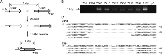
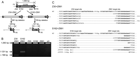
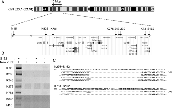
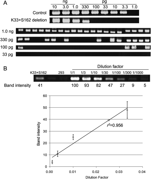
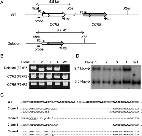
We present a novel approach for generating targeted deletions of genomic segments in human and other eukaryotic cells using engineered zinc finger nucleases (ZFNs). We found that ZFNs designed to target two different sites in a human chromosome could introduce two concurrent DNA double-strand breaks (DSBs) in the chromosome and give rise to targeted deletions of the genomic segment between the two sites. Using this method in human cells, we were able to delete predetermined genomic DNA segments in the range of several-hundred base pairs (bp) to 15 mega-bp at frequencies of 10(-3) to 10(-1). These high frequencies allowed us to isolate clonal populations of cells, in which the target chromosomal segments were deleted, by limiting dilution. Sequence analysis revealed that many of the deletion junctions contained small insertions or deletions and microhomologies, indicative of DNA repair via nonhomologous end-joining. Unlike other genome engineering tools such as recombinases and meganucleases, ZFNs do not require preinsertion of target sites into the genome and allow precise manipulation of endogenous genomic scripts in animal and plant cells. Thus, ZFN-induced genomic deletions should be broadly useful as a novel method in biomedical research, biotechnology, and gene therapy.


Figures

Figure 1.
Figure 1.
ZFN-induced genome deletions at the CCR2 and CCR5 loci. (A) Schematic representation of ZFN-mediated genome deletions. Zigzag lines indicate ZFN target sites. F2 and R5 are PCR primers (arrows) used for the detection of genome deletion events. (B) PCR products corresponding to the 15-kbp genomic DNA deletions in cells treated with ZFNs. p3 is the empty plasmid used as a negative control. (C) DNA sequences of PCR products. PCR products were cloned and sequenced. ZFN target sites are shown in boldface letters. Microhomologies are underlined and inserted bases are shown in italics. Dashes indicate deleted bases. Nonconserved bases at the CCR2 and CCR5 loci are shown in lowercase letters. In cases in which a deletion sequence was detected more than once, the number of occurrences is shown in parentheses. (WT) Wild-type DNA sequence.
Figure 2.
Figure 2.
Use of two ZFNs for targeted genome deletions within the CCR5 locus. (A) Schematic representation of two different deletion events within the CCR5 locus. F5 and R5 are PCR primers (arrows) used for the detection of genome deletion events. (B) PCR products validating ZFN-induced genomic deletions. Approximate sizes of PCR products corresponding to deletion events (199 bp and 331 bp) and to the intact wild-type sequence (1060 bp) are indicated as predicted in A. p3 is the empty plasmid used as a negative control. (C) DNA sequences of PCR products corresponding to deletion events. Symbols are as in Figure 1.
Figure 3.
Figure 3.
Large nested genomic deletions. (A) ZFN target sites on a chromosomal ideogram. Arrows indicate the locations of ZFN target sites on the amplified view of the relevant chromosome 3 region. (B) PCR products validating large nested deletions. S162 and each of the seven new ZFNs were coexpressed in HEK 293 cells. DNA sequences of PCR primers used in this analysis are listed in Supplemental Table 3. (C) DNA sequences of PCR products corresponding to deletion events. Symbols are as in Figure 1.
Figure 4.
Figure 4.
Frequencies of genomic deletions induced by ZFNs. (A) “Digital” PCR analysis. (Upper panel) Genomic DNA isolated from cells cotransfected with plasmids encoding K33 and S162 were serially diluted in a buffer and subjected to PCR analysis in a reaction volume of 10 μL. (Lower panel) At critical dilution points, PCR was performed in 20 aliquots. (B) A standard curve for the estimation of deletion frequencies. A plasmid containing a PCR product corresponding to the 33-kbp genomic deletion induced by ZFNs was serially diluted in a solution containing genomic DNA isolated from HEK 293 cells and the diluted samples were subjected to PCR analysis. Intensities of DNA bands corresponding to the deletion event were measured and plotted against dilution factors. At high values, band intensities reached plateau and thus were excluded when the standard curve was plotted. The open square corresponds to the PCR product from cells treated with ZFNs.
Figure 5.
Figure 5.
Analyses of clonal populations of cells whose genomic segments were deleted by ZFNs. (A) Schematic of CCR2 and CCR5 loci in wild-type cells and in clonal cells whose genomic segments were deleted. The probe (black bar) and the restriction enzyme, XbaI, used for Southern blot analysis are indicated. F2, R2, F5, and R5 are PCR primers (arrows) used for the detection of genome deletion events or the intact CCR2 and CCR5 loci. (B) PCR products validating genomic deletions and the intact CCR2 and CCR5 region. PCR primers are shown in parentheses. Clones 1 to 4 are clonal cells isolated by limiting dilution. (WT) Wild-type cells. (C) DNA sequences of deletion junctions in clonal cells. Symbols are as in Figure 1. (D) Southern blot analysis of clonal cells.

References

    1. Agrawal L, Lu X, Qingwen J, VanHorn-Ali Z, Nicolescu IV, McDermott DH, Murphy PM, Alkhatib G. Role for CCR5Δ32 protein in resistance to R5, R5X4, and X4 human immunodeficiency virus type 1 in primary CD4+ cells. J Virol. 2004;78:2277–2287. - PMC - PubMed
    1. Aten JA, Stap J, Krawczyk PM, van Oven CH, Hoebe RA, Essers J, Kanaar R. Dynamics of DNA double-strand breaks revealed by clustering of damaged chromosome domains. Science. 2004;303:92–95. - PubMed
    1. Bae KH, Kwon YD, Shin HC, Hwang MS, Ryu EH, Park KS, Yang HY, Lee DK, Lee Y, Park J, et al. Human zinc fingers as building blocks in the construction of artificial transcription factors. Nat Biotechnol. 2003;21:275–280. - PubMed
    1. Bibikova M, Golic M, Golic KG, Carroll D. Targeted chromosomal cleavage and mutagenesis in Drosophila using zinc-finger nucleases. Genetics. 2002;161:1169–1175. - PMC - PubMed
    1. Bibikova M, Beumer K, Trautman JK, Carroll D. Enhancing gene targeting with designed zinc finger nucleases. Science. 2003;300:764. - PubMed

Publication types