. 2010 Jan;151(1):174-84.
doi: 10.1210/en.2009-0876. Epub 2009 Dec 1.

Estrogen receptor-alpha as a drug target candidate for preventing lung inflammation

Affiliations

Estrogen receptor-alpha as a drug target candidate for preventing lung inflammation

Elisabetta Vegeto et al. Endocrinology. 2010 Jan.

Abstract

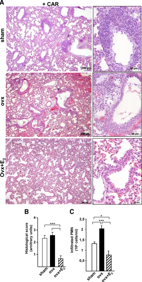
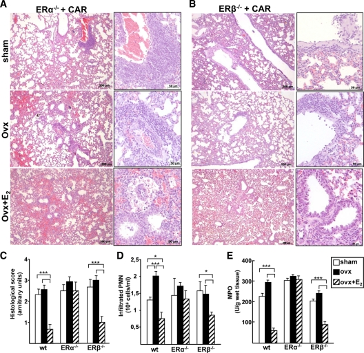
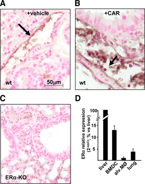
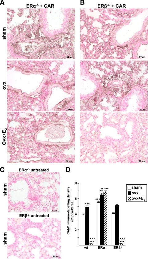
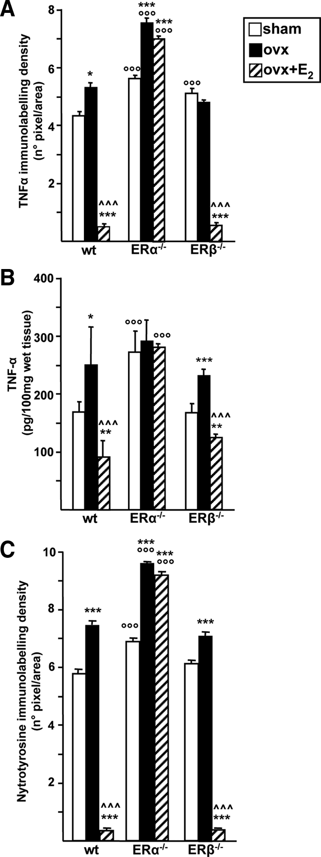
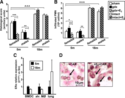
Accumulating evidence shows that estrogens are protective factors in inflammatory lung diseases and are involved in the gender-related incidence of these pathologies. The aim of this study was to identify which estrogen receptor (ER), ER-alpha and/or ER beta, mediates hormone antiinflammatory effects in lung and how gender or aging modify this effect. Acute lung inflammation in wild type, ER alpha or ER beta knockout animals was induced by pleural injection of carrageenan; female mice were used and sham operated, ovariectomized, or ovariectomized and treated with 17beta-estradiol (E(2)) before carrageenan. Our data show that ER alpha, and not ER beta, mediates E(2)-induced reduction of the inflammatory response. By real-time PCR and immunohistochemistry assays, we demonstrate ER alpha expression in the resident and infiltrated inflammatory cells of the lung, in which ER beta could not be detected. In these cells, E(2)-mediated reduction in the expression of inflammatory mediators was also due to ER alpha. In parallel, we observed that female mice were more prone to inflammation as compared with males, suggesting a gender-related difference in lung susceptibility to inflammatory stimuli, whereas the effect of E(2) was similar in the two sexes. Interestingly, aging results in a strong increase in the inflammatory response in both sexes and in the disruption E(2)/ER alpha signaling pathway. In conclusion, our data reveal that E(2) is able to regulate lung inflammation in a gender-unrelated, age-restricted manner. The specific involvement of ER alpha in hormone action opens new ways to identify drug targets that limit the inflammatory component of lung pathologies.


Figures

Figure 1
Figure 1
Estrogen and lung inflammation in female mice. A, Histology of lung sections stained with hematoxylin-eosin obtained from CAR-treated sham, ovx, or ovx females mice treated with E2 (ovx+E2). Representative samples are shown with magnifications on the right. B, As measure of tissue morphology, a histological score was assigned to each animal through the assessment of histological parameters during tissue observation at the microscope, as described in Materials and Methods. Sham, Open boxes; ovx, black boxes; ovx+E2, dashed boxes. C, Pleural exudate was recovered and analyzed for infiltrated PMN cell number; bar legend as in B). Bars represent the average ± sem of all the animals (n = 8), each analyzed in triplicate. *, P < 0.05; ***, P < 0.001.
Figure 2
Figure 2
Role of ER isoforms in lung inflammation. A and B, Histology of lung sections stained with hematoxyline-eosin from ERα- and ERβ-KO mice injected with CAR in the pleural space. Representative samples are shown. C, Histological score. D and E, Pleural exudate was recovered and analyzed (D) for infiltrated PMN cell number and (E) for MPO. Bars represent the average ± sem of all the animals (wt, n = 8; ERα- and ERβ-KO, sham and ovx n = 8; ovx+E2 n = 5), each analyzed in triplicate. *, P < 0.05; ***, P < 0.001.
Figure 3
Figure 3
ERα expression in lung and inflammatory cells. Representative immunohistochemistry images of lung from vehicle (A) and CAR-treated sham (B) or untreated (C) ERα-KO female mice analyzed with an ERα-specific antibody. ERα-positive cells are visible in endothelial cells of the vessel wall (arrow in A) and in round-shaped cells in proximity of vessels (arrow in B). D, Expression levels of the ERα gene was evaluated by real-time PCR on the mRNA extracted from BMDCs, alveolar macrophages (alv. MØ), and lung tissue and represented in relation with ERα mRNA from liver. Each data point represent the average ± sem of samples from three female mice analyzed in triplicate.
Figure 4
Figure 4
ICAM-1 immunolabeling in lung of ER-KO animals. Lung tissue from ERα- (A) or ERβ-KO (B) mice, sham, ovx, or ovx+E2, were analyzed by IHC after CAR injection with specific antibody against ICAM-1. C, IHC of lung sections from ERα- and ERβ-KO untreated mice. Representative immunhistochemistry images are shown. Densitometry evaluation (D) of ICAM-1 immunolabeling in wt, ERα-KO, and ERβ-KO mice. Bar legend as in Fig. 1B. Data are expressed as means of percent of total tissue area ± sem. * vs. sham, ^ vs. ovx; ° vs. corresponding treatment in wt. **, °°, P < 0.01; ***, °°°, P < 0.001.
Figure 5
Figure 5
TNF-α and nytrotyrosine immunolabeling in ER-KO animals. Lung tissue from wt, ERα-KO, or ERβ-KO mice, sham operated, ovx, or ovx+E2 (bar legend as in Fig. 1B) were analyzed after CAR injection with specific antibodies against TNF-α (A) or nytrotyrosine residues (C); densitometry of IHC images were evaluated and data are expressed as means of percent of total tissue area ± sem (B) TNF-α levels were analyzed by immunoenzymatic assay in the pleural exudate. Bars represent the average ± sem of all the animals, each analyzed in triplicate. * vs. sham; ^ vs. ovx; ° vs. corresponding treatment in wt. *, P < 0.05; **, P < 0.01; ***, °°°, ^^^, P < 0.001.
Figure 6
Figure 6
Effect of gender and aging on E2 signaling in lung inflammation. Lung inflammation was induced by CAR injection in female and male mice at the age of 5 (bar legend as in Fig. 1B) or 18 months (either intact or treated with E2), as indicated. Histological score (A) or infiltrated PMN cells numbers (B) are shown; bars represent the average ± sem of all the animals (n = 8), each analyzed in triplicate. * vs. sham; ^ vs. 5-month sham; *, P < 0.05; **, P < 0.01; ***, ^^^, P < 0.001. C, Expression levels of the ERα gene was evaluated by real-time PCR on the mRNA extracted from BMDCs, alveolar macrophages (alv. MØ), and lung tissue from 5-month-old (black bars) and 18-month-old (open bars) female mice. Each data point represents the average ± sem of samples from three mice analyzed in triplicate. D, Representative images of immunohistochemistry assays performed using a specific ERα antibody in lung sections from 18-month-old female mice injected with CAR (left panel) or with E2 before CAR (right panel). A cytoplasmic localization of ERα is observed in PMN cells, independently of E2 administration.

References

    1. Sweezey N, Tchepichev S, Gagnon S, Fertuck K, O'Brodovich H 1998 Female gender hormones regulate mRNA levels and function of the rat lung epithelial Na channel. Am J Physiol 274:C379–C386 - PubMed
    1. Torday JS, Nielsen HC 1987 The sex difference in fetal lung surfactant production. Exp Lung Res 12:1–19 - PubMed
    1. Carey MA, Card JW, Voltz JW, Germolec DR, Korach KS, Zeldin DC 2007 The impact of sex and sex hormones on lung physiology and disease: lessons from animal studies. Am J Physiol Lung Cell Mol Physiol 293:L272–L278 - PubMed
    1. Massaro GD, Mortola JP, Massaro D 1996 Estrogen modulates the dimensions of the lung’s gas-exchange surface area and alveoli in female rats. Am J Physiol 270:L110–L114 - PubMed
    1. Cuzzocrea S, Santagati S, Sautebin L, Mazzon E, Calabrò G, Serraino I, Caputi AP, Maggi A 2000 17β-Estradiol antiinflammatory activity in CAR-induced pleurisy. Endocrinology 141:1455–1463 - PubMed

Publication types

MeSH terms