

Sensory protection of rat muscle spindles following peripheral nerve injury and reinnervation
- PMID: 19952642
- PMCID: PMC3411540
- DOI: 10.1097/PRS.0b013e3181bcee47
Sensory protection of rat muscle spindles following peripheral nerve injury and reinnervation
Abstract
Background: Skeletal muscle structure and function are dependent on intact innervation. Prolonged muscle denervation results in irreversible muscle fiber atrophy, connective tissue hyperplasia, and deterioration of muscle spindles, specialized sensory receptors necessary for proper skeletal muscle function. The protective effect of temporary sensory innervation on denervated muscle, before motor nerve repair, has been shown in the rat. Sensory-protected muscles exhibit less fiber atrophy and connective tissue hyperplasia and maintain greater functional capacity than denervated muscles. The purpose of this study was to determine whether temporary sensory innervation also protects muscle spindles from degeneration.
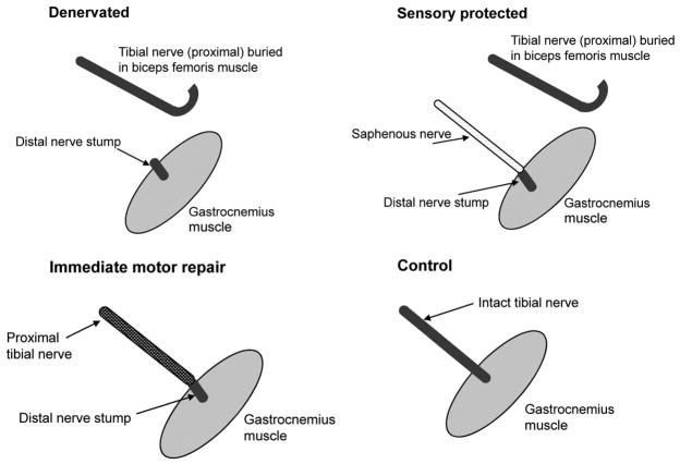
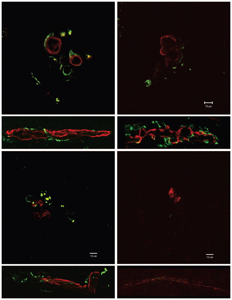
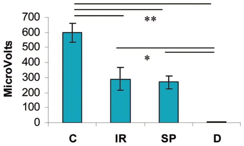
Methods: Rat tibial nerve was transected and repaired with either the saphenous or the original transected nerve. Negative controls remained denervated. After 3 to 6 months, the electrophysiologic response of the nerve to stretch in the rat gastrocnemius muscle was measured (n = 3 per group). After the animals were euthanized, the gastrocnemius muscle was removed, sectioned, stained, and examined for spindle number (n = 3 per group) and morphology (one rat per group). Immunohistochemical assessment of muscle spindle innervation was examined in four additional animals.
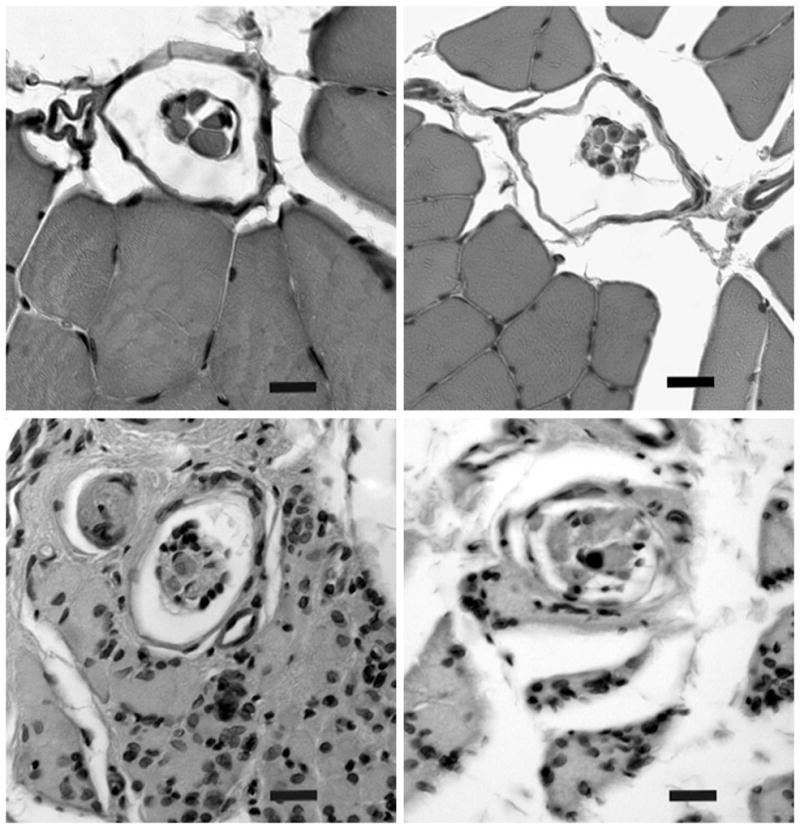
Results: Significant deterioration of muscle spindles was seen in denervated muscle, whereas in muscle reinnervated with the tibial or the saphenous nerve, spindle number and morphology were improved. Histologic and functional evidence of spindle reinnervation by the sensory nerve was obtained.
Conclusion: These findings add to the known means by which motor or sensory nerves exert protective effects on denervated muscle, and further promote the use of sensory protection for improving the outcome after peripheral nerve injury.
Conflict of interest statement
Figures
Similar articles
-
Improved functional recovery of denervated skeletal muscle after temporary sensory nerve innervation.Neuroscience. 2001;103(2):503-10. doi: 10.1016/s0306-4522(00)00577-7. Neuroscience. 2001. PMID: 11246164
-
Sensory nerve cross-anastomosis and electrical muscle stimulation synergistically enhance functional recovery of chronically denervated muscle.Plast Reconstr Surg. 2014 Nov;134(5):736e-745e. doi: 10.1097/PRS.0000000000000599. Plast Reconstr Surg. 2014. PMID: 25347648 Clinical Trial.
-
Early sensory protection in reverse end-to-side neurorrhaphy to improve the functional recovery of chronically denervated muscle in rat: a pilot study.J Neurosurg. 2014 Aug;121(2):415-22. doi: 10.3171/2014.4.JNS131723. Epub 2014 May 30. J Neurosurg. 2014. PMID: 24878291
-
Sensory nerve regeneration and reinnervation in muscle following peripheral nerve injury.Muscle Nerve. 2022 Oct;66(4):384-396. doi: 10.1002/mus.27661. Epub 2022 Jul 2. Muscle Nerve. 2022. PMID: 35779064 Review.
-
Chapter 25: Phototherapy in peripheral nerve injury: effects on muscle preservation and nerve regeneration.Int Rev Neurobiol. 2009;87:445-64. doi: 10.1016/S0074-7742(09)87025-6. Int Rev Neurobiol. 2009. PMID: 19682654 Review.
Cited by
-
Intrafusal-fiber LRP4 for muscle spindle formation and maintenance in adult and aged animals.Nat Commun. 2023 Feb 10;14(1):744. doi: 10.1038/s41467-023-36454-8. Nat Commun. 2023. PMID: 36765071 Free PMC article.
-
Peripheral Nerve Healing: So Near and Yet So Far.Semin Plast Surg. 2021 Aug;35(3):204-210. doi: 10.1055/s-0041-1731630. Epub 2021 Sep 10. Semin Plast Surg. 2021. PMID: 34526869 Free PMC article. Review.
-
Rat whisker movement after facial nerve lesion: evidence for autonomic contraction of skeletal muscle.Neuroscience. 2014 Apr 18;265:9-20. doi: 10.1016/j.neuroscience.2014.01.038. Epub 2014 Jan 28. Neuroscience. 2014. PMID: 24480367 Free PMC article.
-
Morphological differences in skeletal muscle atrophy of rats with motor nerve and/or sensory nerve injury.Neural Regen Res. 2012 Nov 15;7(32):2507-15. doi: 10.3969/j.issn.1673-5374.2012.32.004. Neural Regen Res. 2012. PMID: 25337102 Free PMC article.
-
Bace1 and Neuregulin-1 cooperate to control formation and maintenance of muscle spindles.EMBO J. 2013 Jul 17;32(14):2015-28. doi: 10.1038/emboj.2013.146. Epub 2013 Jun 21. EMBO J. 2013. PMID: 23792428 Free PMC article.
References
-
- Hynes NM, Bain JR, Thoma A, Veltri K, Maguire JA. Preservation of denervated muscle by sensory protection in rats. J Reconstr Microsurg. 1997;13:337–343. - PubMed
-
- Kobayashi J, Mackinnon SE, Watanabe O, et al. The effect of duration of muscle denervation on functional recovery in the rat model. Muscle Nerve. 1997;20:858–866. - PubMed
-
- Bain JR, Veltri KL, Chamberlain D, Fahnestock M. Improved functional recovery of denervated skeletal muscle after temporary sensory nerve innervation. Neuroscience. 2001;103:503–510. - PubMed
-
- Veltri K, Kwiecien JM, Minet W, Fahnestock M, Bain JR. Contribution of the distal nerve sheath to nerve and muscle preservation following denervation and sensory protection. J Reconstr Microsurg. 2005;21:57–70. - PubMed
Publication types
MeSH terms
Grants and funding
LinkOut - more resources
Full Text Sources
