

Vinculin and talin: focus on the myocardium
- PMID: 19952892
- PMCID: PMC2810504
- DOI: 10.2310/JIM.0b013e3181c5e074
Vinculin and talin: focus on the myocardium
Abstract
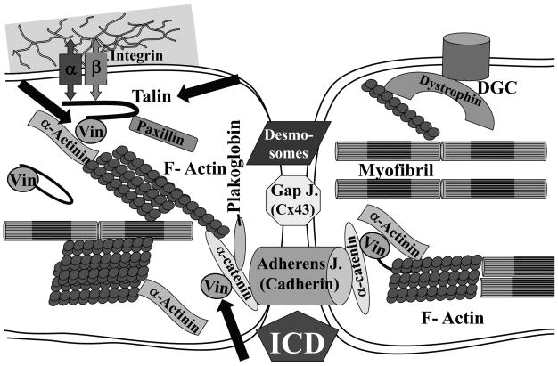
Cardiomyopathy is a heart muscle disease caused by decreased contractility of the ventricles leading to heart failure and premature death. Multiple conditions like ischemic heart disease (atherosclerosis), hypertension, diabetes, viral infection, alcohol abuse, obesity and genetic mutations can lead to cardiomyopathy. Single gene mutations in sarcomeric proteins, Z-disk-associated proteins, membrane/associated proteins, intermediate filaments, calcium cycle proteins as well as in modifier genes have been linked to cardiomyopathy. Clinical practice guidelines have been formulated by the American Heart Association and the Heart Failure Association of America on how to genetically evaluate patients with cardiomyopathy. To illustrate the concept that alterations in genes cause cardiovascular disease, this review will focus on two membrane-associated proteins, vinculin and talin. We will discuss the general function of vinculin/metavinulin as well as talin1 and talin2, with emphasis on what is understood about their role in the cardiac myocyte and in whole heart.
Figures
Similar articles
-
Vinculin, talin and focal adhesions.J Muscle Res Cell Motil. 1996 Feb;17(1):1-5. doi: 10.1007/BF00140319. J Muscle Res Cell Motil. 1996. PMID: 8740427 Review. No abstract available.
-
Integrin connections to the cytoskeleton through talin and vinculin.Biochem Soc Trans. 2008 Apr;36(Pt 2):235-9. doi: 10.1042/BST0360235. Biochem Soc Trans. 2008. PMID: 18363566 Review.
-
The viscoelasticity of entangled actin networks: the influence of defects and modulation by talin and vinculin.Eur Biophys J. 1993;22(5):309-21. doi: 10.1007/BF00213554. Eur Biophys J. 1993. PMID: 8112218
-
Loss of mouse cardiomyocyte talin-1 and talin-2 leads to β-1 integrin reduction, costameric instability, and dilated cardiomyopathy.Proc Natl Acad Sci U S A. 2017 Jul 25;114(30):E6250-E6259. doi: 10.1073/pnas.1701416114. Epub 2017 Jul 11. Proc Natl Acad Sci U S A. 2017. PMID: 28698364 Free PMC article.
-
Intermolecular versus intramolecular interactions of the vinculin binding site 33 of talin.Protein Sci. 2011 Aug;20(8):1471-6. doi: 10.1002/pro.671. Protein Sci. 2011. PMID: 21648001 Free PMC article.
Cited by
-
Passive myocardial mechanical properties: meaning, measurement, models.Biophys Rev. 2021 Oct 13;13(5):587-610. doi: 10.1007/s12551-021-00838-1. eCollection 2021 Oct. Biophys Rev. 2021. PMID: 34765043 Free PMC article. Review.
-
Myofilament dysfunction in diastolic heart failure.Heart Fail Rev. 2024 Jan;29(1):79-93. doi: 10.1007/s10741-023-10352-z. Epub 2023 Oct 14. Heart Fail Rev. 2024. PMID: 37837495 Free PMC article. Review.
-
Vinculin at the heart of aging.Ann Transl Med. 2017 Feb;5(3):62. doi: 10.21037/atm.2017.01.65. Ann Transl Med. 2017. PMID: 28251141 Free PMC article. No abstract available.
-
The intercalated disc: a mechanosensing signalling node in cardiomyopathy.Biophys Rev. 2020 Aug;12(4):931-946. doi: 10.1007/s12551-020-00737-x. Epub 2020 Jul 13. Biophys Rev. 2020. PMID: 32661904 Free PMC article. Review.
-
Mechanotransduction in cardiac hypertrophy and failure.Circ Res. 2015 Apr 10;116(8):1462-1476. doi: 10.1161/CIRCRESAHA.116.304937. Circ Res. 2015. PMID: 25858069 Free PMC article. Review.
References
-
- Ahmad F, Seidman JG, Seidman CE. The genetic basis for cardiac remodeling. Annu Rev Genomics Hum Genet. 2005;6:185–216. - PubMed
-
- Alcalai R, Seidman JG, Seidman CE. Genetic basis of hypertrophic cardiomyopathy: from bench to the clinics. J Cardiovasc Electrophysiol. 2008;19:104–110. - PubMed
-
- Ho CY, Seidman CE. A contemporary approach to hypertrophic cardiomyopathy. Circulation. 2006;113:e858–e862. - PubMed
-
- Sabatine MS, Seidman JG, Seidman CE. Cardiovascular genomics. Circulation. 2006;113:e450–e455. - PubMed
-
- Hershberger RE, Lindenfeld J, Mestroni L, et al. Genetic evaluation of cardiomyopathy--a Heart Failure Society of America practice guideline. J Card Fail. 2009;15:83–97. - PubMed
Publication types
MeSH terms
Substances
Grants and funding
LinkOut - more resources
Full Text Sources
