. 2010 Jan;33(1):16-29.
doi: 10.1097/CJI.0b013e3181b0b146.

The marginating-pulmonary immune compartment in rats: characteristics of continuous inflammation and activated NK cells

Affiliations

The marginating-pulmonary immune compartment in rats: characteristics of continuous inflammation and activated NK cells

Rivka Melamed et al. J Immunother. 2010 Jan.

Abstract

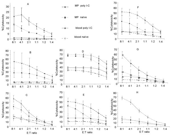
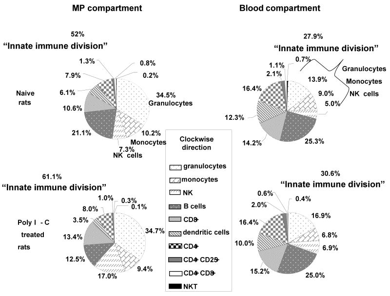
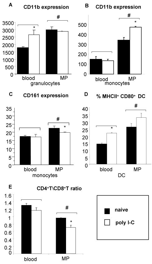
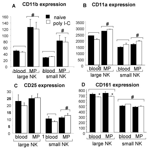
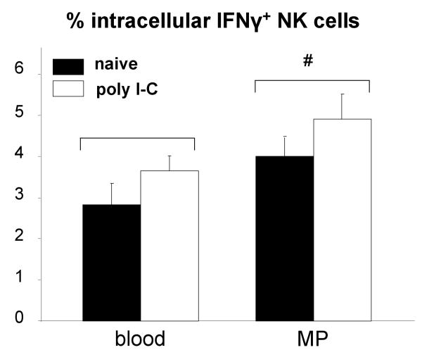
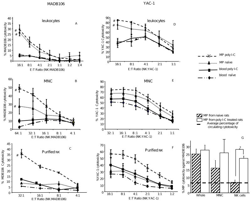
A significant role has been indicated for cellular immunity in controlling circulating cancer cells, but most autologous tumor cells seem resistant, in vitro, to natural killer cell (NKC) and cytotoxic T lymphocytes cytotoxicity. Addressing this apparent contradiction, we recently identified a unique leukocyte population, marginating-pulmonary (MP)-leukocytes, which exhibit potent natural killer (NK) cytotoxicity. Here, we characterize the MP-compartment in naive and immunostimulated rats, and assessed its cytotoxicity against "NK-resistant" tumors cells. Animals were treated with poly I-C (3x0.2 mg/kg) or saline, and circulating-leukocytes and MP-leukocytes were collected and analyzed in terms of cellular composition, cellular activation markers, and NK cytotoxicity of leukocytes and purified NKCs. Compared with circulating-leukocytes, MP-leukocytes showed greater proportion of granulocytes, monocytes, NKCs, and large NKCs; higher expression of activation and adhesion markers (CD25, CD11a, CD11b, and NKR-P1, IFN-gamma); and elevated NK cytotoxicity of leukocytes and purified NKCs against several syngeneic and xenogeneic NK-resistant target cells (from both F344 and BDX inbred rats). In immunostimulated animals (treated with poly I-C), but not in naive animals, purified NKCs from the MP-compartment showed markedly superior cytotoxicity, suggesting that poly I-C immunostimulation uniquely affect MP-NKCs, and that in naive animals other MP-leukocytes support NK cytotoxicity. Overall, the results suggest that the MP-compartment is characterized by a continuous activated inflammatory microenvironment uniquely affected by immunostimulation. If similarly potent MP-NKCs exist in patients, then circulating autologous tumor cells that are considered "NK-resistant" could actually be controlled by MP-NKCs. Innate immunity may assume greater role in controlling malignant spread, especially after immunostimulation.


Figures

Fig.1
Fig.1. Higher MP-NK cytotoxicity against syngeneic, allogeneic, and xenogeneic cell lines of the F344 and BDX rat strains
MP-leukocytes from F344 (Fig.1A-D) and BDX's (Fig.1E-H) rats exhibited marked and significant higher NK cytotoxicity against various syngeneic, allogeneic, and xenogeneic tumor cells compare to leukocytes from the circulation (P < 0.005, for each tumor line). NK cytotoxicity in F344 rats was tested against the syngeneic MADB106 (Fig.1A) and CRNK-16 (Fig.1B), and the xenogeneic P815 (Fig.1C) and YAC-1 (Fig.1D) tumor cell lines. NK cytotoxicity in BDX rats was examined against the syngeneic 73AS (Fig.1E), AS-14 (Fig.1F), and ASML (Fig.1G), and the allogeneic MADB106 (Fig.1H) tumor lines. Cytotoxicity levels are presented in all figures using the same NK:target cell ratio. The lysis of all syngeneic tumor lines by circulating leukocytes was very low (Fig 1A, B, E, F, and G). Poly I-C immunostimulation significantly increased MP-NK, but not citculating-NK cytotoxicity, against all tumor lines (excluding the xenogeneic YAC-1, in which cytotoxicity increased in both compartments). Data are expressed as mean ± SEM.
Fig.2
Fig.2. Different leukocyte subsets composition in the MP-compartment vs. the circulation
The MP-compartment exhibited a markedly different cellular composition than the circulation: the “innate immune division”, including granulocytes, monocytes, and NK cells, constituted 52% (± 1.52%) of the MP-leukocytes population, compared to 27.9% (± 0.66%) in the circulation (p < 0.05). Contrary, the T cell subpopulations, including CD4+, CD4+CD8+ T cells, and regulatory (CD4+ CD25+) T cells, as well as DC, exhibited lower percentage in the MP compartment compared to the circulation (P < = 0.005). Following poly I-C administration, the proportion of the “innate immune division” in the MP-compartment further increased, reaching 61.1% (±2.56%) (P < 0.05), and also increased in the circulation to only 21% (±1.48 %) (P < 0.05).
Fig.3
Fig.3. Phenotypes of an active inflammatory state in the MP-leukocytes population
Compared to the circulation, MP-granulocytes exhibited higher expression levels of CD11b (Fig.3A) (P < 0.05), and MP-monocytes expressed higher levels of CD11b and NKR-P1 (Fig.3B-C). MP-DCs contained a higher percentage of mature DCs compared to circulating DCs, as indicated by higher expression levels of the CD80+MHC-II+ cells (Fig.3D), and poly I-C further increased it in both compartments. The CD4+T/CD8+T ratio was significantly reduced in the MP-compartment compared to the circulation, and poly I-C further decreased it in both compartments (Fig.3E). # indicate significant difference between the MP compartment and the blood compartment, and * indicates significant difference within a compartment, between poly I-C and naive animals. Data are expressed as mean ± SEM.
Fig.4
Fig.4. NK cells' number, percentage, and size, is greater in the MP-compartment
Leukocytes were labeled by two fluorescent markers to identify CD161 (FL-1, for NK cells) and CD5 (FL-2, for T cells). Together with FSC and SSC, small NK cells and large NK were identified, and are circled in panel A (T cells, granulocytes, and plastic beads are not marked in the figure). In the 4 scattergrams of panel A, FSC by FL-1 are presented for circulating and MP-leukocytes, treated or not with poly I-C. See that NK cells can be clearly categorized into two distinct subpopulations: large NK cells (larger than 18 μm) and small NK cells (Fig 4A). The total number of NK cells per MP-leukocytes and per ml blood increased following poly I-C treatment (p < 0.05 for both), but significantly more in the MP-compartment (4.2 ± 2 fold vs. 1.5 ± 1.0) (p < 0.05) (Fig.4B). MP-NK cells contained a three-fold higher percentage of large NK cells than circulating NK cells (30% vs. 10%, respectively) (p < 0.05) (Fig 4C), and poly I-C did not affect this ratio in neither compartments. # indicate significant difference between the MP compartment and the blood compartment, and * indicates significant difference within a compartment, between poly I-C and naive animals. Data are expressed as mean ± SEM.
Fig.5
Fig.5. Different expression levels of activation and adhesion surface markers on small and large NK cells in the two immune compartments
Compared to small NK cells, large NK cells were characterized by significantly higher expression levels of the adhesion molecules CD11b (Mac-1) (Fig.5A) and CD11a (LFA-1) (Fig.5B), and of CD25 (IL-2 receptor) (Fig.5C) and CD161 (NKR-P1) (Fig.5D), (p < 0.05 for all). These differences were evident in both compartments, and in both poly I-C and saline treated rats. In addition, MP-NK cells also exhibit significantly and markedly higher expression levels of CD11b and CD11a compared to circulating-NK cells (Fig 5A-B). # indicate significant difference between the MP compartment and the blood compartment, and * indicates significant difference within a compartment, between poly I-C and naive animals. Data are expressed as mean ± SEM
Fig.6
Fig.6. Higher intracellular interferon gamma in MP-NK cells
Intracellular IFN-γ was expressed by a significantly higher percentage of MP-NK cells, compared to circulating NK cells (#) (p < 0.05), and poly I-C further increased this percentage in both compartments (P < 0.05 across compartments) (Fig.6). Data are expressed as mean ± SEM.
Fig.7
Fig.7. NK cytotoxicity against MADB106 and YAC-1 target cells along increasing levels of NK cell purification
A & D. Circulating and MP-leukocytes: MP-leukocytes exhibited marked and significant NK cytotoxicity compared to circulating-leukocytes against MADB106 (7A) and YAC-1 target cells (7D) (P < 0.05 for each). Poly I-C had no significant effects. B & E. Circulating and MP-MNC: MP-MNC exhibited marked and significant higher NK cytotoxicity compared to circulating-MNC against the MADB106 (Fig.7B) and YAC-1 (7E) target cells (P < 0.05 for both). Poly I-C increased NK cytotoxicity, but significantly so only in MP-MNC cytotoxicity against the MADB106. C & F. Purified circulating and MP-NK cells: No differences were evident between MP and circulating purified NK cells from naïve rats. However, poly I-C increased NK cytotoxicity only in the MP compartment, in a marked and significant manner (Fig 7C and 7 F) (P < 0.05), as indicated by a significant interaction between the compartment and the effects of poly I-C (P < 0.05). In all panels, data are expressed as mean ± SEM. G. Comparison of MADB106 cytotoxicity by MP-NK cells between the three purification levels at a common E:T ratio (16:1) (Taken from A-C). Cytotoxicity levels of circulating NK cells of all purification levels are below the dashed line (5-12%). The more we enriched the part of NKCs (from all leukocytes, to MNC, to purified NK cells) the more MP-NK cell cytotoxicity decreased, and the smaller the differences between the cytotoxicity of the two immune compartments became. However, pre-exposure to poly I-C maintained the levels of MP-NK cytotoxicity and thus the difference between the two compartments.
Fig.8
Fig.8. The effect of in vivo NK depletion on controlling MADB106 metastases and on in vitro MADB106 lysis
Selective elimination of NK cells markedly increased lung tumor retention (A) and lung tumor metastases (B) in both poly I-C treated and untreated rats (n = 40, P<0.001). (Notice the logarithmic scale). In vitro MP-NK activity against MADB106 target cells was significantly greater in control and in IL-12 or poly I-C treated rats compared to NK-depleted rats. Depletion of NK cells markedly decreased NK activity to a similar level in IL-12, poly I-C, and vehicle treated groups (C) (n = 53, P<0.001). This, together with the inability of circulating and other populations of NK cells to lyse MADB106, suggest the role of MP-NK cells in controlling MADB106 cells in vivo. Data are expressed asmean ± SEM.

Similar articles

Cited by

References

    1. Shakhar G, Ben-Eliyahu S. Potential prophylactic measures against postoperative immunosuppression: could they reduce recurrence rates in oncological patients? Ann Surg Oncol. 2003;10(8):972–92. - PubMed
    1. Smyth MJ, Godfrey DI, Trapani JA. A fresh look at tumor immunosurveillance and immunotherapy. Nat Immunol. 2001;2(4):293–9. - PubMed
    1. Moretta L, et al. Human natural killer cells: Molecular mechanisms controlling NK cell activation and tumor cell lysis. Immunol Lett. 2005;100(1):7–13. - PubMed
    1. Brittenden J, et al. Natural killer cells and cancer. Cancer. 1996;77(7):1226–43. - PubMed
    1. Cerwenka A, Lanier LL. Natural killer cells, viruses and cancer. Nat Rev Immunol. 2001;1(1):41–9. - PubMed

Publication types