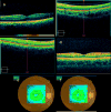
Comparison of spectral/Fourier domain optical coherence tomography instruments for assessment of normal macular thickness
- PMID: 19952997
- PMCID: PMC2819609
- DOI: 10.1097/IAE.0b013e3181bd2c3b
Comparison of spectral/Fourier domain optical coherence tomography instruments for assessment of normal macular thickness
Abstract
Purpose: The purpose of this study was to report normal macular thickness measurements and assess reproducibility of retinal thickness measurements acquired by a time-domain optical coherence tomography (OCT) (Stratus, Carl Zeiss Meditec, Inc., Dublin, CA) and three commercially available spectral/Fourier domain OCT instruments (Cirrus HD-OCT, Carl Zeiss Meditec, Inc.; RTVue-100, Optovue, Inc., Fremont, CA; 3D OCT-1000, Topcon, Inc., Paramus, NJ).
Methods: Forty randomly selected eyes of 40 normal, healthy volunteers were imaged. Subjects were scanned twice during 1 visit and a subset of 25 was scanned again within 8 weeks. Retinal thickness measurements were automatically generated by OCT software and recorded after manual correction. Regression and Bland-Altman plots were used to compare agreement between instruments. Reproducibility was analyzed by using intraclass correlation coefficients, and incidence of artifacts was determined.
Results: Macular thickness measurements were found to have high reproducibility across all instruments with intraclass correlation coefficients values ranging 84.8% to 94.9% for Stratus OCT, 92.6% to 97.3% for Cirrus Cube, 76.4% to 93.7% for RTVue MM5, 61.1% to 96.8% for MM6, 93.1% to 97.9% for 3D OCT-1000 radial, and 31.5% to 97.5% for 3D macular scans. Incidence of artifacts was higher in spectral/Fourier domain instruments, ranging from 28.75% to 53.16%, compared with 17.46% in Stratus OCT. No significant age or sex trends were found in the measurements.
Conclusion: Commercial spectral/Fourier domain OCT instruments provide higher speed and axial resolution than the Stratus OCT, although they vary greatly in scanning protocols and are currently limited in their analysis functions. Further development of segmentation algorithms and quantitative features are needed to assist clinicians in objective use of these newer instruments to manage diseases.
Figures


References
-
- Puliafito CA, Hee MR, Lin CP, et al. Imaging of macular diseases with optical coherence tomography. Ophthalmology. 1995;102(2):217–229. - PubMed
-
- Hee MR, Puliafito CA, Wong C, et al. Quantitative assessment of macular edema with optical coherence tomography. Arch Ophthalmol. 1995;113(8):1019–1029. - PubMed
-
- Schuman JS, Hee MR, Puliafito CA, et al. Quantification of nerve fiber layer thickness in normal and glaucomatous eyes using optical coherence tomography. Arch Ophthalmol. 1995;113(5):586–596. - PubMed
-
- de Boer JF, Cense B, Park BH, et al. Improved signal-to-noise ratio in spectral-domain compared with time-domain optical coherence tomography. Opt Lett. 2003;28(21):2067–2069. - PubMed
Publication types
MeSH terms
Grants and funding
LinkOut - more resources
Full Text Sources
Other Literature Sources
Medical

