Clinical Trial
. 2010 Mar;18(3):643-50.
doi: 10.1038/mt.2009.277. Epub 2009 Dec 1.

Gene therapy for Leber's congenital amaurosis is safe and effective through 1.5 years after vector administration

Affiliations
Clinical Trial

Gene therapy for Leber's congenital amaurosis is safe and effective through 1.5 years after vector administration

Francesca Simonelli et al. Mol Ther. 2010 Mar.

Abstract

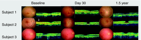
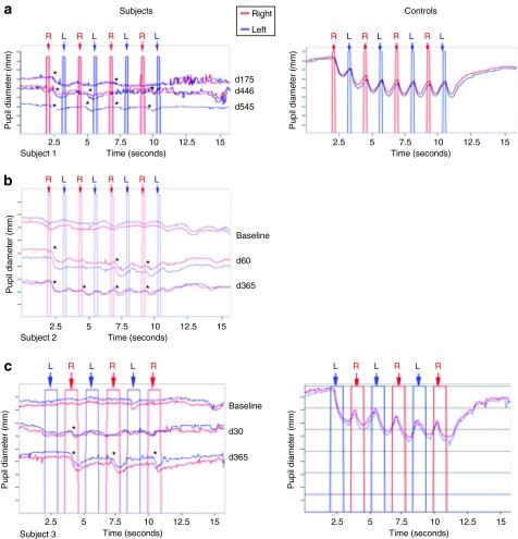
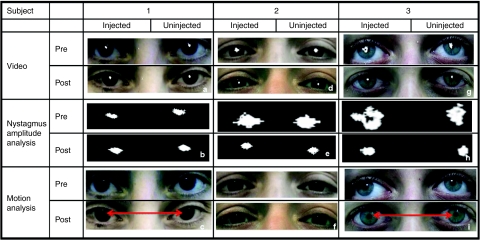
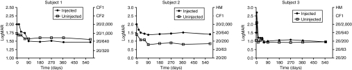
The safety and efficacy of gene therapy for inherited retinal diseases is being tested in humans affected with Leber's congenital amaurosis (LCA), an autosomal recessive blinding disease. Three independent studies have provided evidence that the subretinal administration of adeno-associated viral (AAV) vectors encoding RPE65 in patients affected with LCA2 due to mutations in the RPE65 gene, is safe and, in some cases, results in efficacy. We evaluated the long-term safety and efficacy (global effects on retinal/visual function) resulting from subretinal administration of AAV2-hRPE65v2. Both the safety and the efficacy noted at early timepoints persist through at least 1.5 years after injection in the three LCA2 patients enrolled in the low dose cohort of our trial. A transient rise in neutralizing antibodies to AAV capsid was observed but there was no humoral response to RPE65 protein. The persistence of functional amelioration suggests that AAV-mediated gene transfer to the human retina does not elicit immunological responses which cause significant loss of transduced cells. The persistence of physiologic effect supports the possibility that gene therapy may influence LCA2 disease progression. The safety of the intervention and the stability of the improvement in visual and retinal function in these subjects support the use of AAV-mediated gene augmentation therapy for treatment of inherited retinal diseases.


Figures

<b>Figure 1</b>
Figure 1
Fundus photographs and corresponding optical coherence tomography images through the fovea are shown for all three subjects at baseline, day 30, and 1.5 years after gene delivery. In subject 2, the epiretinal membrane is visible at baseline (arrow). A full-thickness macular hole is apparent on day 30 and 1.5 years. The adjacent retina remains attached.
<b>Figure 2</b>
Figure 2
Representative long-term follow-up pupillometry results in the three subjects. The pupillary light reflexes after dark adaptation are shown as a response to alternating stimulation of the right eye and the left eye. The curves represent the diameter of the pupils; the right pupil is slightly higher than the left pupil in each trace where two pupils are shown. The following pupil traces were displaced vertically to facilitate comparisons: subject 1 day 545, subject 2 baseline, day 365, day 545; subject 3 baseline, subject 3 day 365, subject 3 day 545. The average pupil diameters immediately prior to the first light exposure in the series were as follows: subject 1, 6.4 mm for both pupils at day 175; 5.6 (right), 5.28 (left) at day 446, and 4.9 (right) at day 545; subject 2: 7.1 for both pupils at baseline, 7.8 mm (right) and 7.1 mm (left) pupil at day 60; 4.7 mm for both pupils at day 365 and 4.96 mm (left) pupil at day 545; subject 3: 6.6 mm for both pupils at baseline; 6.3 (right) and 6.1 (left) at day 30; 5.8 (right), 5.4 at day 305 and 4.56 mm at day 545. Alternating stimulation with light of 10.0 lux, 10.0 lux, and 0.04 lux, respectively for subjects 1, 2, and 3. Timepoints (days) with respect to injection are indicated to the right of the traces. Results for test 1 [exposure of eyes to alternating brief (0.2 seconds) flashes of light] are shown for subjects 1 and 2; results for test 2 [exposure of eyes to alternating long (1.0 seconds) flashes of light] are shown for subject 3. Alternating stimuli were presented 2 seconds after the recording was initiated.
<b>Figure 3</b>
Figure 3
Pre- and postinjection nystagmus analysis. (a,b) Frames from pre- and postinjection video recordings in subject 1 with motion paths of the pupils superimposed. Motion of each eye is shown by indicating the location of the center of the pupil over 100 sequential frames (total of 4 seconds) isolated from the video recordings. In b, the motion path is isolated. In c, pre- and postinjection measurements in subject 1 show reduction in interpupillary distance with concomitant changes in the corneal light reflexes (arrowheads). Similar analyses are shown for (d–f) subject 2 and (g–i). Analyses show a significant decrease in binocular amplitude of nystagmus in primary position in subjects 2 and 3 after injection and a smaller decrease in amplitude in subject 1 after injection. There is also a reduction in interpupillary distance in (i) subject 3.
<b>Figure 4</b>
Figure 4
Visual acuity (LogMAR on the left y axis and Snellen value on the right y axis) over time after gene delivery. Baseline is indicated as day 0; gene delivery (no visual acuity measurement) is on day 1.

Comment in

  • The eyes have it.
    Brenner M. Brenner M. Mol Ther. 2010 Mar;18(3):451-2. doi: 10.1038/mt.2010.12. Mol Ther. 2010. PMID: 20195259 Free PMC article. No abstract available.

References

    1. Cremers FP, van den Hurk JA., and , den Hollander AI. Molecular genetics of Leber congenital amaurosis. Hum Mol Genet. 2002;11:1169–1176. - PubMed
    1. Fulton AB, Hansen RM., and , Mayer DL. Vision in Leber congenital amaurosis. Arch Ophthalmol. 1996;114:698–703. - PubMed
    1. Aleman TS, Jacobson SG, Chico JD, Scott ML, Cheung AY, Windsor EA, et al. Impairment of the transient pupillary light reflex in Rpe65(−/−) mice and humans with leber congenital amaurosis. Invest Ophthalmol Vis Sci. 2004;45:1259–1271. - PubMed
    1. Simonelli F, Ziviello C, Testa F, Rossi S, Fazzi E, Bianchi PE, et al. Clinical and molecular genetics of Leber's congenital amaurosis: a multicenter study of Italian patients. Invest Ophthalmol Vis Sci. 2007;48:4284–4290. - PubMed
    1. den Hollander AI, Roepman R, Koenekoop RK., and , Cremers FP. Leber congenital amaurosis: genes, proteins and disease mechanisms. Prog Retin Eye Res. 2008;27:391–419. - PubMed

Publication types