

Disease-associated extracellular matrix suppresses osteoblastic differentiation of human periodontal ligament cells via MMP-1
- PMID: 19953231
- PMCID: PMC3152822
- DOI: 10.1007/s00223-009-9321-z
Disease-associated extracellular matrix suppresses osteoblastic differentiation of human periodontal ligament cells via MMP-1
Abstract
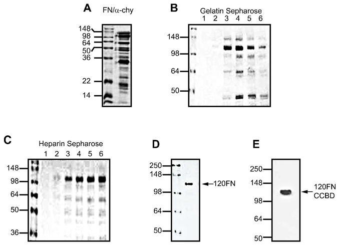
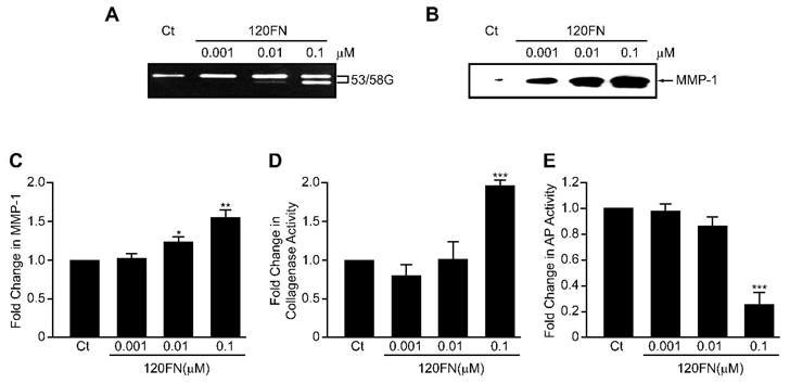
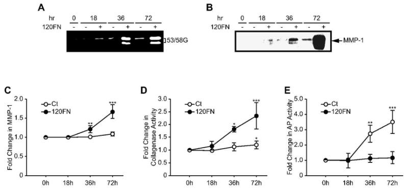
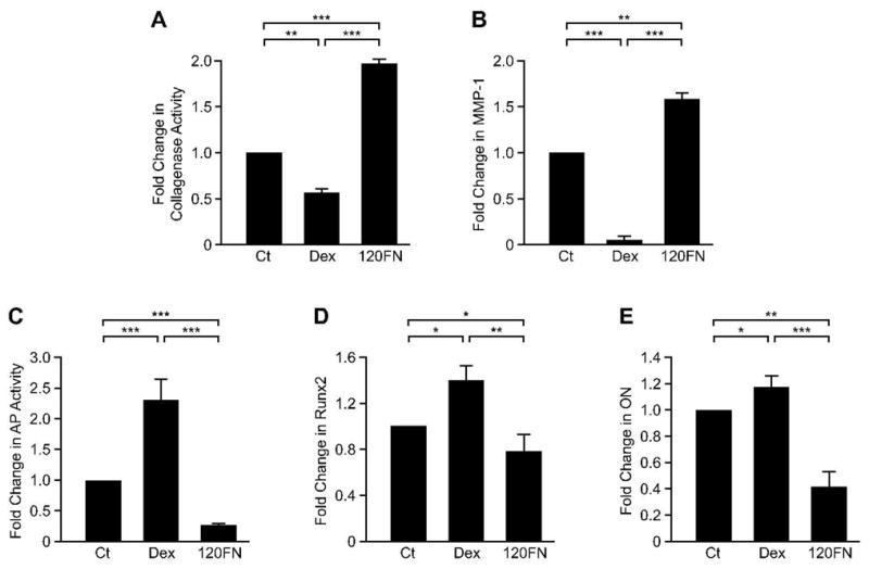
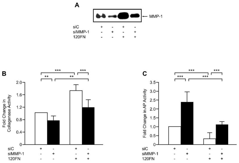
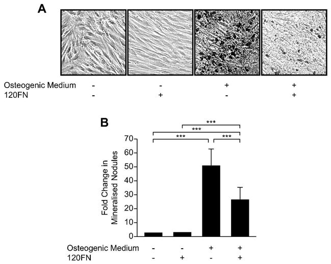
Fibronectin (FN) fragments found in chronic inflammatory diseases, including periodontal disease and arthritis, may contribute to tissue destruction in part via induction of matrix metalloproteinases (MMPs). We previously showed that the 120-kDa FN fragment containing the central cell binding domain (120FN) dose dependently induces MMP-1 (collagenase-1) in human periodontal ligament (PDL) cells, whereas intact FN did not elicit this response. Recently, we found that an increase in MMP-1 expression is accompanied by a decreased osteoblastic phenotype in PDL cells. We hypothesized that 120FN inhibits osteoblastic differentiation of PDL cells by inducing MMP-1. Effects of increasing concentrations of 120FN on MMP-1 expression and on osteoblastic markers were assessed in cultured PDL cells using Western blotting, qRT-PCR, and collagen degradation and alkaline phosphatase (AP) activity assays. The 120FN dose dependently increased MMP-1 expression and activity, concomitant with a decrease in AP activity. The increase in collagenase activity was largely attributed to increased MMP-1 expression. Concurrent with the decrease in AP activity, the 120FN reduced baseline and dexamethasone-induced gene expression of specific osteoblastic markers, Runx2 and osteonectin, and diminished mineralized nodule formation. Finally, siRNA inhibition of 120FN-induced MMP-1 reduced collagenase expression and rescued the AP phenotype to baseline levels. These findings suggest that disease-associated 120FN, in addition to having direct effects on tissue destruction by upregulating MMPs, could contribute to disease progression by impeding osteoblastic differentiation of osteogenic PDL cells and, consequently, diminish bone regeneration.
Figures
Similar articles
-
MMP-1 (collagenase-1) and MMP-13 (collagenase-3) differentially regulate markers of osteoblastic differentiation in osteogenic cells.Matrix Biol. 2008 Oct;27(8):682-92. doi: 10.1016/j.matbio.2008.07.005. Epub 2008 Aug 5. Matrix Biol. 2008. PMID: 18755271 Free PMC article.
-
Dexamethasone's enhancement of osteoblastic markers in human periodontal ligament cells is associated with inhibition of collagenase expression.Bone. 2007 Jan;40(1):93-104. doi: 10.1016/j.bone.2006.07.003. Epub 2006 Aug 24. Bone. 2007. PMID: 16934542
-
Ascorbic acid induces collagenase-1 in human periodontal ligament cells but not in MC3T3-E1 osteoblast-like cells: potential association between collagenase expression and changes in alkaline phosphatase phenotype.J Bone Miner Res. 2003 Jan;18(1):67-77. doi: 10.1359/jbmr.2003.18.1.67. J Bone Miner Res. 2003. PMID: 12510807
-
Fibronectin and fibronectin fragments modulate the expression of proteinases and proteinase inhibitors in human periodontal ligament cells.Matrix Biol. 1996 Sep;15(4):251-61. doi: 10.1016/s0945-053x(96)90116-x. Matrix Biol. 1996. PMID: 8892225
-
Matrix metalloproteinases in periodontal tissue remodelling.Matrix Suppl. 1992;1:352-62. Matrix Suppl. 1992. PMID: 1480060 Review.
Cited by
-
Characterization of ligamentum flavum hypertrophy based on m6A RNA methylation modification and the immune microenvironment.Am J Transl Res. 2022 Dec 15;14(12):8800-8827. eCollection 2022. Am J Transl Res. 2022. PMID: 36628248 Free PMC article.
-
Shedding of NG2 by MMP-13 attenuates anoikis.DNA Cell Biol. 2014 Dec;33(12):854-62. doi: 10.1089/dna.2014.2399. DNA Cell Biol. 2014. PMID: 25166220 Free PMC article.
-
The Treponema denticola chymotrypsin-like protease dentilisin induces matrix metalloproteinase-2-dependent fibronectin fragmentation in periodontal ligament cells.Infect Immun. 2011 Feb;79(2):806-11. doi: 10.1128/IAI.01001-10. Epub 2010 Nov 29. Infect Immun. 2011. PMID: 21115719 Free PMC article.
-
Treponema denticola upregulates MMP-2 activation in periodontal ligament cells: interplay between epigenetics and periodontal infection.Arch Oral Biol. 2014 Oct;59(10):1056-64. doi: 10.1016/j.archoralbio.2014.06.003. Epub 2014 Jun 14. Arch Oral Biol. 2014. PMID: 24973519 Free PMC article.
-
Treponema denticola increases MMP-2 expression and activation in the periodontium via reversible DNA and histone modifications.Cell Microbiol. 2018 Apr;20(4):10.1111/cmi.12815. doi: 10.1111/cmi.12815. Epub 2018 Jan 8. Cell Microbiol. 2018. PMID: 29205773 Free PMC article.
References
-
- Barkalow FJ, Schwarzbauer JE. Localization of the major heparin-binding site in fibronectin. J Biol Chem. 1991;266:7812–7818. - PubMed
-
- Hahn LH, Yamada KM. Isolation and biological characterization of active fragments of the adhesive glycoprotein fibronectin. Cell. 1979;18:1043–1051. - PubMed
-
- Clemmensen I, Andersen RB. Different molecular forms of fibronectin in rheumatoid synovial fluid. Arthritis Rheum. 1982;25:25–31. - PubMed
Publication types
MeSH terms
Substances
Grants and funding
LinkOut - more resources
Full Text Sources
Medical
Miscellaneous
