Review
. 2010 Jan;334(1-2):117-24.
doi: 10.1007/s11010-009-0328-6. Epub 2009 Dec 2.

Mg2+/Ca2+ cation binding cycle of guanylyl cyclase activating proteins (GCAPs): role in regulation of photoreceptor guanylyl cyclase

Affiliations
Review

Mg2+/Ca2+ cation binding cycle of guanylyl cyclase activating proteins (GCAPs): role in regulation of photoreceptor guanylyl cyclase

Alexander M Dizhoor et al. Mol Cell Biochem. 2010 Jan.

Abstract

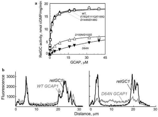
Photon absorption by photoreceptors activates hydrolysis of cGMP, which shuts down cGMP-gated channels and decreases free Ca(2+) concentrations in outer segment. Suppression of Ca(2+) influx through the cGMP channel by light activates retinal guanylyl cyclase through guanylyl cyclase activating proteins (GCAPs) and thus expedites photoreceptors recovery from excitation and restores their light sensitivity. GCAP1 and GCAP2, two ubiquitous among vertebrate species isoforms of GCAPs that activate retGC during rod response to light, are myristoylated Ca(2+)/Mg(2+)-binding proteins of the EF-hand superfamily. They consist of one non-metal binding EF-hand-like domain and three other EF-hands, each capable of binding Ca(2+) and Mg(2+). In the metal binding EF-hands of GCAP1, different point mutations can selectively block binding of Ca(2+) or both Ca(2+) and Mg(2+) altogether. Activation of retGC at low Ca(2+) (light adaptation) or its inhibition at high Ca(2+) (dark adaptation) follows a cycle of Ca(2+)/Mg(2+) exchange in GCAPs, rather than release of Ca(2+) and its binding by apo-GCAPs. The Mg(2+) binding in two of the EF-hands controls docking of GCAP1 with retGC1 in the conditions of light adaptation and is essential for activation of retGC. Mg(2+) binding in a C-terminal EF-hand contributes to neither retGC1 docking with the cyclase nor its subsequent activation in the light, but is specifically required for switching the cyclase off in the conditions of dark adaptation by binding Ca(2+). The Mg(2+)/Ca(2+) exchange in GCAP1 and 2 operates within different range of intracellular Ca(2+) concentrations and provides a two-step activation of the cyclase during rod recovery.


Figures

Fig. 1
Fig. 1
Ca2+ feedback and cGMP synthesis in photoreceptors. Explanations are provided in the text
Fig. 2
Fig. 2
Three-dimensional structure of GCAP1 [41] and GCAP2 [40]. GCAPs comprise two semi-globular halves formed by two pairs of EF-hand structures (EF-1+EF-2 and EF-3+EF-4), numbered beginning from the N-terminus in primary structure. Spheres represent divalent cation bound in EF-hand loops. The loop in the first EF-hand domain lacks some key consensus side chain residues required for coordinating the metal ions. The arrows indicate the positions of two mutations associated with congenital blindness, Y99C and E155G. Other explanations are given in the text
Fig. 3
Fig. 3
Mg2+ binding makes GCAP1 into RetGC activator in the conditions of light adaptation whereas Ca2+ binding is required to decelerate the cyclase at high Ca2+ concentrations typical for dark-adapted rods (from [26, 30]). a Mutations in 12-amino acid EF-had loops of each of EF-hands 2, 3, and 4 that can disable either Ca2+ or both Ca2+ and Mg2+ binding [26] in EF-hand loops of bovine GCAP1. b Activation of recombinant RetGC1 by mutant forms GCAP1 was assayed in vitro at different free Ca2+ concentrations in the presence of Ca/Mg/EGTA buffer maintaining Mg2+ at near 5 mM level. Wild-type GCAP1 (open circle) produces maximal retGC activity below 200 nM free Ca2+ but decelerates it as Ca2+ rises; inactivation of only Ca2+ binding turns GCAP1 into a Ca2+-insensitive Mg2+-bound activator of RetGC (filled square), whereas mutations preventing both Ca2+ and Mg2+ binding turn GCAP1 into an apo-form and completely block activation of RetGC (filled triangle). c Cation binding cycle in GCAP1 as it undergoes transitions between the RetGC activator and inhibitor states, driven by light versus dark exposure of photoreceptors. Other explanations are given in the text
Fig. 4
Fig. 4
Effects of Mg2+ binding prevention in individual EF-hands (reproduced from [35] with minor modifications). a Inactivation of Mg2+ binding in EF-hands 2 and 3, but not in EF-hand 4 inhibits RetGC1 activation by GCAP1. Mutations blocking only Ca2+ binding in all three EF-hands (open circle) or inactivation of both Ca2+ and Mg2+ binding in EF-hand 4 (filled triangle) do not alter RetGC activation by GCAP1 (wt, open square). Mutation blocking both Ca2+ and Mg2+ binding in EF-hand 3 (open triangle) and especially EF-hand 2 (filled inverted triangle) suppress activation of RetGC1; measured at constant 5 mM free Mg2+ and <20 nM free Ca2+. For other details see reference [35]. b Inactivation of Mg2+ binding in EF-hand 2 suppresses docking of GCAP1-GFP with RetGC1 in cultured cells (modified from [35]). RetGC1 was co-expressed in transfected HEK293 cells with GFP-tagged GCAP1, either wild type (left) or its D64N mutant (right), incapable of binding Mg2+ in EF-hand 2. Distribution of the GFP tag green fluorescence and anti-RetGC1 red immunofluorescence was recorded across each cell. For other details see [35]
Fig. 5
Fig. 5
Ca2+ and Mg2+ sensitivity of RetGC regulation by GCAP1 and GCAP2 in rod outer segment (ROS) membranes (reproduced from [33]). a, b ROS membranes were depleted from the endogenous GCAPs by multiple hypotonic extractions, reconstituted with recombinant GCAP1 (a) or GCAP2 (b), and total RetGC activity in ROS was assayed as a function of free Ca2+ in the presence of 0.5 mM (open square), 1 mM (filled triangle), 2 mM (filled circle), and 5 mM (inverted filled triangle) free Mg2+; basal RetGC activity is shown at 0.5, 1, 2, and 5 mM Mg2+ (filled square, filled diamond, open circle, and open diamond, respectively) in the ROS membranes without GCAPs added. c The [Ca]1/2 values for RetGC inhibition by GCAP1 and GCAP2 increase proportionally with the free Mg2+ concentration, but their relative difference remains unchanged
Fig. 6
Fig. 6
Two-step activation model of retGC by GCAP1 and GCAP2 (reproduced from [54]). a Neither GCAP1 nor GCAP2 activate retGC in the dark when the Ca2+ concentration in rods is high. After illumination, as soon as Ca2+ starts to fall, GCAP1, which has the lower affinity for Ca2+ of the two GCAPs, converts into a Mg2+-bound state and activates retGC. This activation alone is not fully sufficient to achieve the normal, physiological kinetics of photoresponse recovery and light adaptation. Any fall in Ca2+ below 100 nM stimulates Ca2+/Mg2+ exchange to occur in GCAP2, which then provides additional activation of retGC. At present, it remains unclear whether or not each GCAP acts through a particular retGC isozyme or whether both GCAPs can directly or via an allosteric mechanisms compete for the same isozyme(s). b Slowed recovery of the GCAP2−/− single-photon response. The mean, fractional single-photon response from the knockout rods (gray trace) was similar in size to that from the wild-type rods (black trace), but the recovery was slower. The divergence of the two traces late in the rising phase suggests that GCAP2 normally activates retGC at this time. See [54], for the details

Similar articles

Cited by

References

    1. Pugh EN, Jr, Duda T, Sitaramayya A, Sharma RK. Photoreceptor guanylate cyclases: a review. Biosci Rep. 1997;17:429–473. - PubMed
    1. Pugh EN, Jr, Nikonov S, Lamb TD. Molecular mechanisms of vertebrate photoreceptor light adaptation. Curr Opin Neurobiol. 1999;9:410–418. - PubMed
    1. Nakatani K, Chen C, Yau KW, Koutalos Y. Calcium and phototransduction. Adv Exp Med Biol. 2002;514:1–20. - PubMed
    1. Burns ME, Mendez A, Chen J, Baylor DA. Dynamics of cyclic GMP synthesis in retinal rods. Neuron. 2002;36:81–91. - PubMed
    1. Hodgkin AL, Nunn BJ. Control of light-sensitive current in salamander rods. J Physiol. 1988;403:439–471. - PMC - PubMed

Publication types

LinkOut - more resources