

Reliability and precision of pseudo-continuous arterial spin labeling perfusion MRI on 3.0 T and comparison with 15O-water PET in elderly subjects at risk for Alzheimer's disease
- PMID: 19953503
- PMCID: PMC2843795
- DOI: 10.1002/nbm.1462
Reliability and precision of pseudo-continuous arterial spin labeling perfusion MRI on 3.0 T and comparison with 15O-water PET in elderly subjects at risk for Alzheimer's disease
Abstract
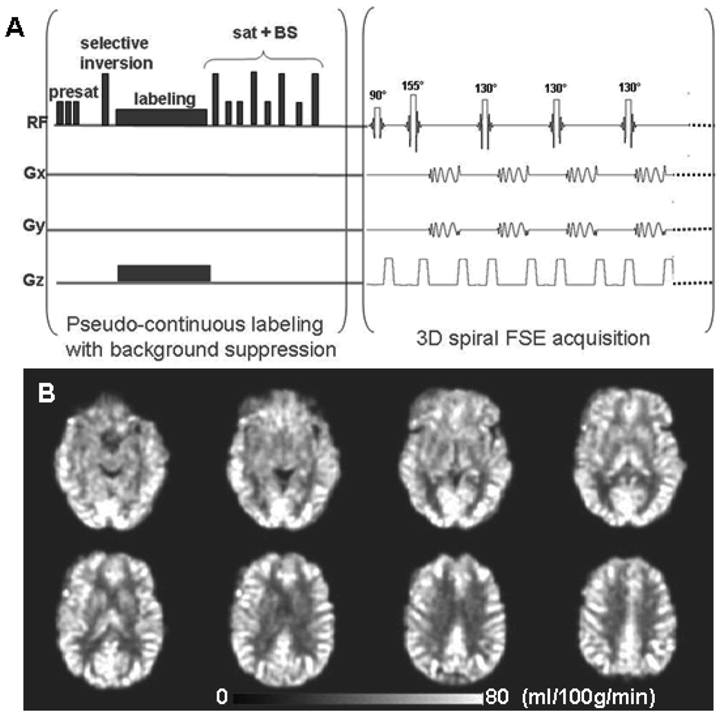
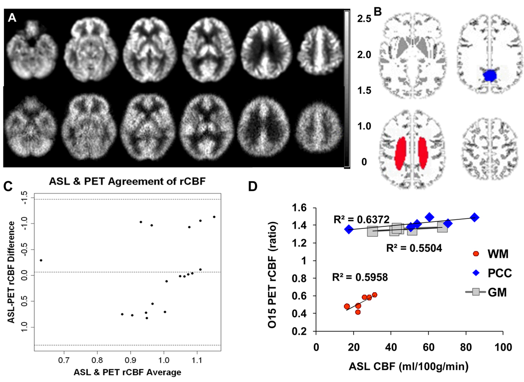
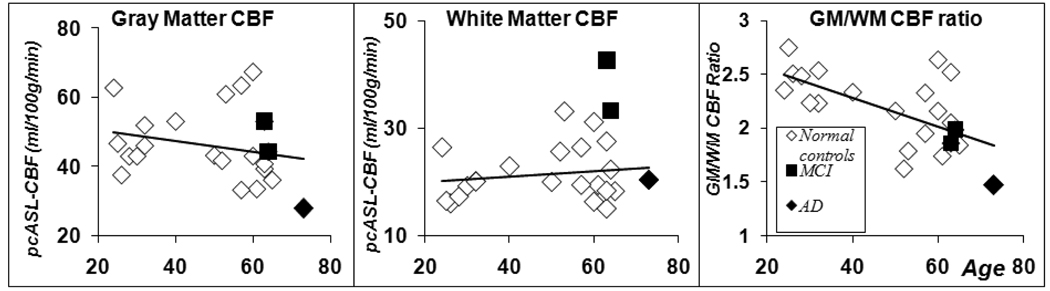
Arterial spin labeling (ASL) offers MRI measurement of cerebral blood flow (CBF) in vivo, and may offer clinical diagnostic utility in populations such as those with early Alzheimer's disease (AD). In the current study, we investigated the reliability and precision of a pseudo-continuous ASL (pcASL) sequence that was performed two or three times within one hour on eight young normal control subjects, and 14 elderly subjects including 11 with normal cognition, one with AD and two with Mild Cognitive Impairment (MCI). Six of these elderly subjects including one AD, two MCIs and three controls also received (15)O-water positron emission tomography (PET) scans 2 h before their pcASL MR scan. The instrumental reliability of pcASL was evaluated with the intraclass correlation coefficient (ICC). The ICCs were greater than 0.90 in pcASL global perfusion measurements for both the young and the elderly groups. The cross-modality perfusion imaging comparison yielded very good global and regional agreement in global gray matter and the posterior cingulate cortex. Significant negative correlation was found between age and the gray/white matter perfusion ratio (r = -0.62, p < 0.002). The AD and MCI patients showed the lowest gray/white matter perfusion ratio among all the subjects. The data suggest that pcASL provides a reliable whole brain CBF measurement in young and elderly adults whose results converge with those obtained with the traditional (15)O-water PET perfusion imaging method. pcASL perfusion MRI offers an alternative method for non-invasive in vivo examination of early pathophysiological changes in AD.
2009 John Wiley & Sons, Ltd.
Figures
References
-
- Huang C, Wahlund LO, Almkvist O, Elehu D, Svensson L, Jonsson T, Winblad B, Julin P. Voxel- and VOI-based analysis of SPECT CBF in relation to clinical and psychological heterogeneity of mild cognitive impairment. Neuroimage. 2003;19(3):1137–1144. - PubMed
-
- Ishii K, Sasaki M, Yamaji S, Sakamoto S, Kitagaki H, Mori E. Demonstration of decreased posterior cingulate perfusion in mild Alzheimer's disease by means of H215O positron emission tomography. European journal of nuclear medicine. 1997;24(6):670–673. - PubMed
-
- Alsop DC, Detre JA, Grossman M. Assessment of cerebral blood flow in Alzheimer's disease by spin-labeled magnetic resonance imaging. Ann Neurol. 2000;47(1):93–100. - PubMed
Publication types
MeSH terms
Substances
Grants and funding
LinkOut - more resources
Full Text Sources
Medical
