. 2009 Dec 29;48(51):12252-64.
doi: 10.1021/bi901498v.

Characterization of [4Fe-4S]-containing and cluster-free forms of Streptomyces WhiD

Affiliations

Characterization of [4Fe-4S]-containing and cluster-free forms of Streptomyces WhiD

Jason C Crack et al. Biochemistry. .

Abstract

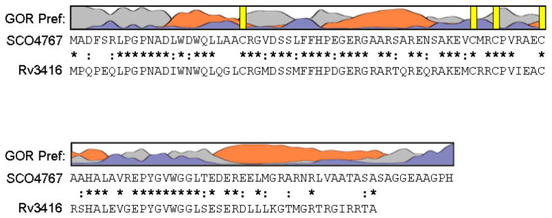
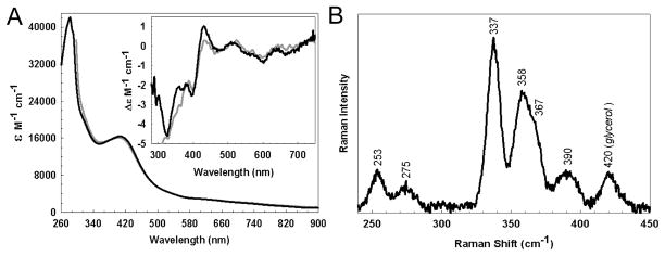
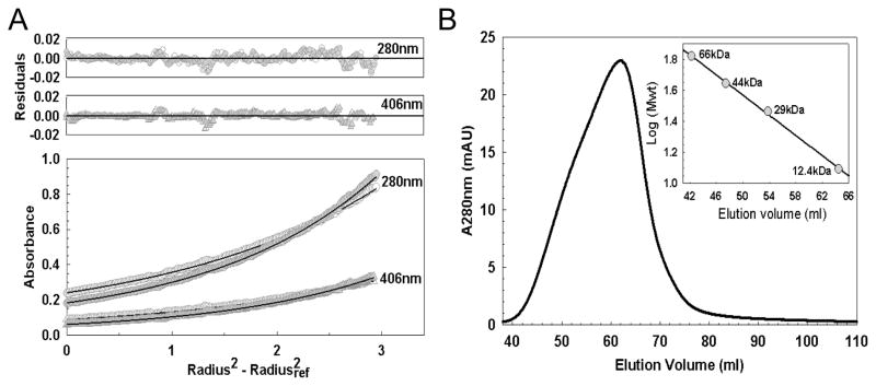
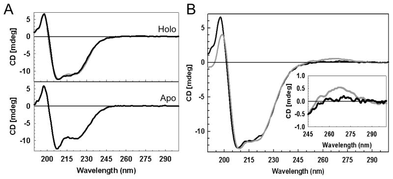
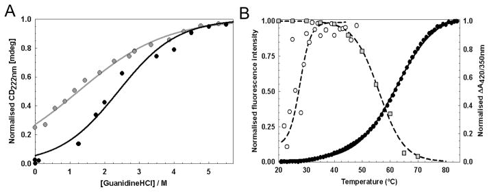
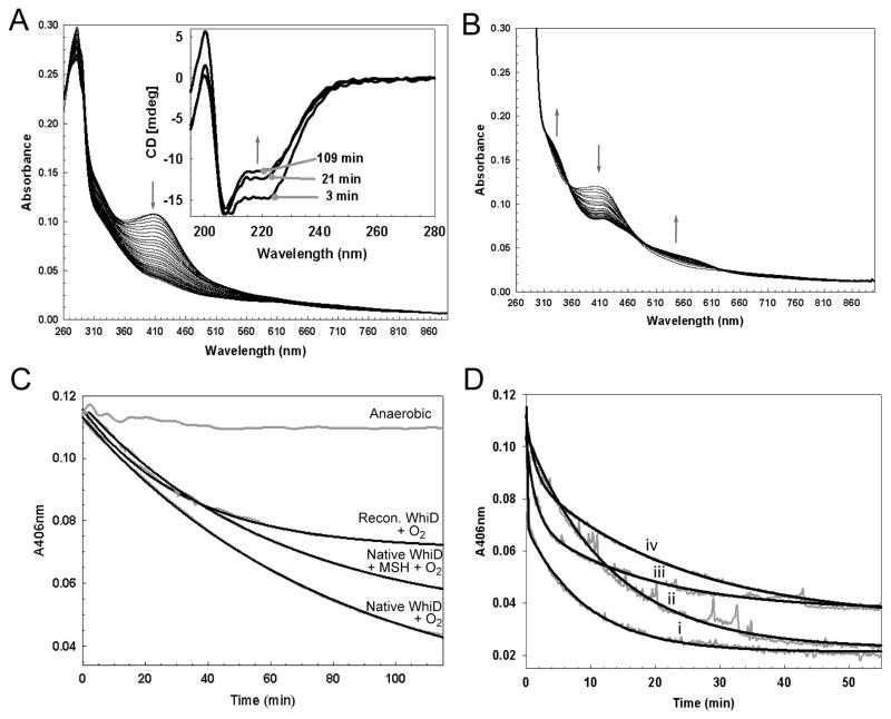
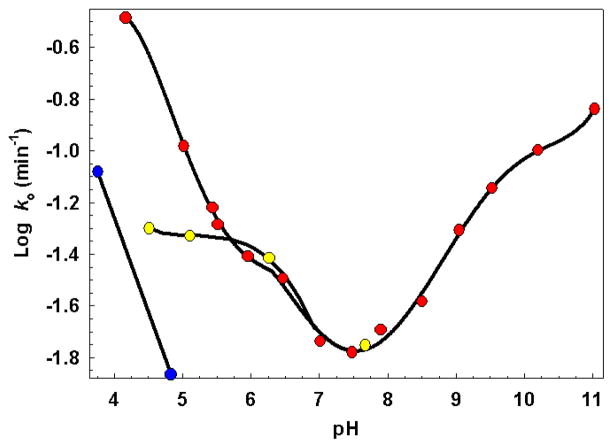
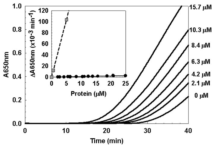
WhiD, a member of the WhiB-like (Wbl) family of iron-sulfur proteins found exclusively within the actinomycetes, is required for the late stages of sporulation in Streptomyces coelicolor. Like all other Wbl proteins, WhiD has not so far been purified in a soluble form that contains a significant amount of cluster, and characterization has relied on cluster-reconstituted protein. Thus, a major goal in Wbl research is to obtain and characterize native protein containing iron-sulfur clusters. Here we report the analysis of S. coelicolor WhiD purified anaerobically from Escherichia coli as a soluble protein containing a single [4Fe-4S](2+) cluster ligated by four cysteines. Upon exposure to oxygen, spectral features associated with the [4Fe-4S] cluster were lost in a slow reaction that unusually yielded apo-WhiD directly without significant concentrations of cluster intermediates. This process was found to be highly pH dependent with an optimal stability observed between pH 7.0 and pH 8.0. Low molecular weight thiols, including a mycothiol analogue and thioredoxin, exerted a small but significant protective effect against WhiD cluster loss, an activity that could be of physiological importance. [4Fe-4S](2+) WhiD was found to react much more rapidly with superoxide than with either oxygen or hydrogen peroxide, which may also be of physiological significance. Loss of the [4Fe-4S] cluster to form apoprotein destabilized the protein fold significantly but did not lead to complete unfolding. Finally, apo-WhiD exhibited negligible activity in an insulin-based disulfide reductase assay, demonstrating that it does not function as a general protein disulfide reductase.


Figures

Figure 1
Figure 1. Sequence analysis of WhiD (SCO4767)
The secondary structure of WhiD was predicted using GOR IV (73). The resulting prediction indicated approximately 42% α-helix, 55% coil and 3% beta strand. Preference for α-helix is indicated by orange, β-sheet by blue and coil by grey. Vertical yellow bars indicate the position of cysteine residues. Sequence alignment of WhiD (SCO4767) and WhiB3 (Rv3416) from M. tuberculosis H37Rv is shown. Conserved residues are indicated by a star, conservatively substituted residues are indicated by a colon.
Figure 2
Figure 2. Spectroscopic characterisation of native WhiD
(A) Optical absorption spectrum and Inset CD spectrum of native WhiD (396 μM [4Fe-4S], 1mm pathlength) in 50 mM Tris 250 mM NaCl 5% (v/v) glycerol, pH 8.0. Extinction coefficients relate to the [4Fe-4S]2+ concentration. The equivalent spectrum arising from in vitro refolded/reconstituted WhiD (dashed gray line) is shown for comparison. (B) Low temperature (17 K) resonance Raman spectrum of native WhiD (0.3 mM) in the Fe-S stretching region using 457.9 nm laser excitation. The spectrum is the sum of 100 scans with each scan involving photon counting for 1 s every 0.5 cm−1 with 6 cm−1 spectral resolution.
Figure 3
Figure 3. Association state of holo-WhiD
(A) Equilibrium analytical ultracentrifugation of native-WhiD (21.5 μM) in 50 mM Tris-HCl 250 mM NaCl pH 8.0. The distribution of WhiD was tracked via protein and cofactor absorbancies at 280 nm (circle) and 406 nm (triangle), respectively, in the presence (gray filled) and absence (white filled) of 7 mM DTT, as indicated. Data were fitted to a single component model (black lines, see Experimental procedures) giving a molecular mass of 14.6 kDa and 15.3 kDa in the presence or absence of 7 mM DTT, respectively. Fit residuals are shown above the main plot. (B) Gel filtration chromatogram of native WhiD (450 μM) in 50 mM Tris-HCl 250 mM NaCl pH 8.0. WhiD eluted at a volume corresponding to a molecular mass of ~15 kDa. Inset: Standard calibration curve for the Sephacryl 100HR column.
Figure 4
Figure 4. Secondary structure analysis of WhiD
(A) Far UV CD spectra of native (upper pane) and apo- (lower pane) WhiD (14.1 μM protein) in 50 mM Tris 250 mM NaCl 5% (v/v) glycerol, pH 8.0. A simulation of the native WhiD spectrum generated by the K2D (48) spectral deconvolution programme (gray line, upper pane) is plotted for comparison. (B) Comparison of far UV CD spectra of refolded/reconstituted (gray line) and native (black line) [4Fe-4S] WhiD. Inset is an expanded view of the near-UV CD region.
Figure 5
Figure 5. Conformational stability of WhiD
(A) Chemical denaturation of apo- (gray circles) and native-WhiD (black circles) (14.1 μM protein) in 50 mM Tris-HCl, 25 mM NaCl, pH 8.0 with GdnHCl between 0 to 6 M. Fraction of unfolded protein (as normalised intensity at 222 nm) is plotted as a function of GdnHCl concentration. Apo-WhiD displayed a non sigmoidal transition suggesting it is partly unfolded in the absence of denaturant. Assuming apo-WhiD is ~25% unfolded at zero denaturant, both apo- and native-WhiD fitted to a two-state unfolding model (solid lines, see main text). (B) Thermal denaturation of apo- (open circles) and native- WhiD (filled circles) followed by tryptophan fluorescence changes. The thermal stability of the iron-sulfur cluster was followed by plotting the ratio of A420 nm to A350 nm (gray squares, see Table 1). Lines represent fits of the data obtained using the Cary Eclipse Bio Software or Origin (Microcal) to obtain Tm and ΔGunfold values.
Figure 6
Figure 6. Reactivity of the [4Fe-4S] cluster
(A) Absorption spectra of native-WhiD (~7 μM [4Fe-4S]) following addition to an aerobic buffer (20 mM Tris HCl, 20 mM Mes, 20 mM BisTrisPropane, 100 mM NaCl 5% Glycerol (v/v), pH 8.0); spectra were recorded every 5 min. Inset, Changes in the far UV CD spectra of native-WhiD in response to O2. Spectra were recorded 3, 21 and 109 min after exposure to O2, as indicated. Arrows indicate the direction of movement of spectral features. (B) Absorption spectra of refolded/reconstituted WhiD (~7 μM [4Fe-4S]) following addition to an aerobic buffer; spectra were recorded every 5 min. (C) Plots of A406 nm versus time for anaerobic and aerobic samples of native-WhiD and of refolded/reconstituted WhiD. The data (gray lines) were fitted to a single exponential function (black lines), yielding pseudo first order rate constants. The buffer contained ~234 μM dissolved O2. (D) Plots of ΔA406 nm versus time for the reaction of native WhiD (~7 μM [4Fe-4S]) with i) KO2 (see Supporting Figure S2 for time dependent UV-visible spectra), ii) KO2 plus 780 units superoxide dismutase, iii) KO2 plus 1267 units catalase and iv) KO2 plus superoxide dismutase and catalase, 780 and 1267 units, respectively. The data (gray lines) were fitted to single (ii and iv) or double (i and iii) exponential functions (black lines), yielding pseudo first order rate constants. The buffer contained ~43 μM dissolved superoxide ion (see Experimental procedures). Note that the data are noisy because the experiments were performed with continuous stirring using a spectrometer with a fibre optic link to the anaerobic chamber.
Figure 7
Figure 7. pH sensitivity of the [4Fe-4S] cluster
Pseudo first order rate constants for cluster loss from native-WhiD (7 μM [4Fe-4S]) in aerobic (red circles), aerobic in the presence of methyl mycothiol (yellow circles) and anaerobic (blue circles) buffer are plotted as a function of pH. Solid black lines, derived from polynomial fits, are intended only to indicate overall trends. The buffer was 20 mM Tris HCl, 20 mM Mes, 20 mM BisTrisPropane, 100 mM NaCl 5% Glycerol (v/v), pH as indicated. At pH ≤ 5.5 or ≥ 9.0, 20 mM sodium citrate or 20 mM sodium carbonate, respectively, was added to increase buffer capacity (see Experimental procedures). Solutions contained ~234 μM dissolved O2, 2 mM GSH and 0.5 mM methyl mycothiol as required.
Figure 8
Figure 8. General protein disulfide reductase activity of apo-WhiD
Insulin disulfide reduction assay of native apo-WhiD measured as an increase in A650 nm due to the precipitation of insulin caused by the reduction of the soluble oxidized form. The concentration of WhiD in each experiment is indicated. Inset is a plot of the relative reductase activity as a function of protein concentration for native apo-WhiD and E. coli thioredoxin.

References

    1. den Hengst CD, Buttner MJ. Redox control in actinobacteria. Biochim Biophys Acta. 2008;1780:1201–1216. - PubMed
    1. Soliveri JA, Gomez J, Bishai WR, Chater KF. Multiple paralogous genes related to the Streptomyces coelicolor developmental regulatory gene whiB are present in Streptomyces and other actinomycetes. Microbiology. 2000;146(Pt 2):333–343. - PubMed
    1. Molle V, Palframan WJ, Findlay KC, Buttner MJ. WhiD and WhiB, homologous proteins required for different stages of sporulation in Streptomyces coelicolor A3(2) J Bacteriol. 2000;182:1286–1295. - PMC - PubMed
    1. Davis NK, Chater KF. The Streptomyces coelicolor whiB gene encodes a small transcription factor-like protein dispensable for growth but essential for sporulation. Mol Gen Genet. 1992;232:351–358. - PubMed
    1. Morris RP, Nguyen L, Gatfield J, Visconti K, Nguyen K, Schnappinger D, Ehrt S, Liu Y, Heifets L, Pieters J, Schoolnik G, Thompson CJ. Ancestral antibiotic resistance in Mycobacterium tuberculosis. Proc Natl Acad Sci USA. 2005;102:12200–12205. - PMC - PubMed

Publication types

MeSH terms

Substances