. 2010 Jan 27;426(1):13-7.
doi: 10.1042/BJ20091321.

Rapid turnover of mitochondrial uncoupling protein 3

Affiliations

Rapid turnover of mitochondrial uncoupling protein 3

Vian Azzu et al. Biochem J. .

Abstract

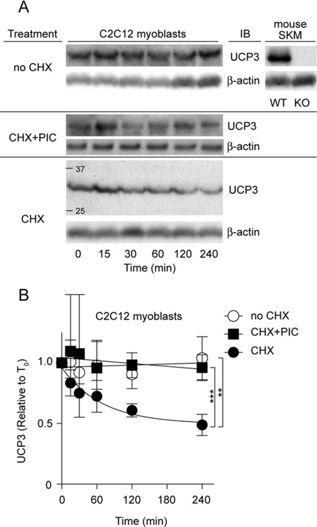
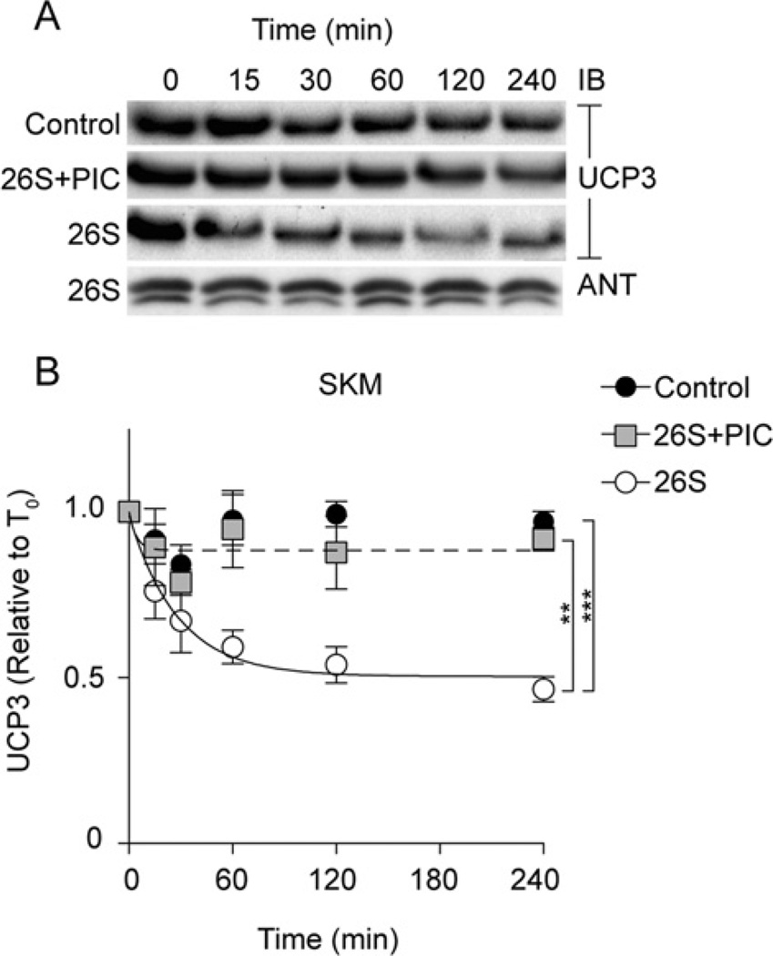
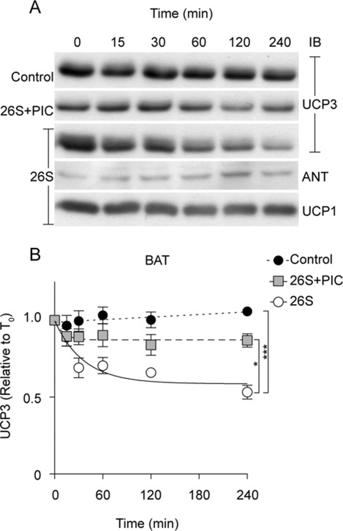
UCP3 (uncoupling protein 3) and its homologues UCP2 and UCP1 are regulators of mitochondrial function. UCP2 is known to have a short half-life of approx. 1 h, owing to its rapid degradation by the cytosolic 26S proteasome, whereas UCP1 is turned over much more slowly by mitochondrial autophagy. In the present study we investigate whether UCP3 also has a short half-life, and whether the proteasome is involved in UCP3 degradation. UCP3 half-life was examined in the mouse C2C12 myoblast cell line by inhibiting protein synthesis with cycloheximide and monitoring UCP3 protein levels by immunoblot analysis. We show that UCP3 has a short half-life of 0.5-4 h. Rapid degradation was prevented by a cocktail of proteasome inhibitors, supporting a proteasomal mechanism for turnover. In addition, this phenotype is recapitulated in vitro: UCP3 was degraded in mitochondria isolated from rat skeletal muscle or brown adipose tissue with a half-life of 0.5-4 h, but only in the presence of a purified 26S proteasomal fraction. This in vitro proteolysis was also sensitive to proteasome inhibition. This phenotype is in direct contrast with the related proteins UCP1 and the adenine nucleotide translocase, which have long half-lives. Therefore UCP3 is turned over rapidly in multiple cell types in a proteasome-dependent manner.


Figures

Figure 1
Figure 1. Proteasome inhibitors block UCP3 degradation in C2C12 cells
(A) C2C12 cells were treated with 10 µg/ml cycloheximide (CHX) at time zero. Cells were harvested at the times shown, separated by SDS/PAGE (1×10⁵ cells/lane) and immunoblotted (IB) for UCP3 (and β-actin). Immunoblot signals were UCP3-specific, being present in skeletal muscle mitochondria from WT, but not KO, mice. One representative blot is shown. (B) UCP3 degradation kinetics based on immunoblot signals. Where indicated, cells were pre-incubated with PIC (containing 10 µM MG132, 10 µM lactacystin and 30 µM PI-1) for 2 h before addition of CHX. Values are means±S.E.M. (n =4), corrected for loading (β-actin). Statistical significance was determined by repeated-measures ANOVA (comparison of matching non-zero time points) with Dunnett’s post-hoc testing (**P < 0.01, ***P < 0.001).
Figure 2
Figure 2. Reconstitution of UCP3 degradation in skeletal muscle mitochondria
The reconstituted system (see the Experimental section) consisted of 1 mg/ml isolated rat skeletal muscle mitochondria in STE buffer (pH 7.4) incubated at 37°C with an ATP-regeneration system (0.5 mM ATP, 10 mM phosphocreatine and 20 units/ml creatine kinase), ubiquitination fractions (50 µg of ubiquitin, 1.6 µg of fraction 1 and 1.6 µg of fraction 2), 2 µg of 26S proteasome fraction, 20 mM succinate and 50 µM PIC as indicated. Aliquots were removed at the time points shown. (A) Proteins (25 µg of protein/lane) were separated by SDS/PAGE and immunoblotted (IB) for UCP3 and ANT. One representative blot is shown. (B) UCP3 degradation kinetics based on immunoblot signals. Values are means±S.E.M. (n =3–5), normalized to ANT. Statistical significance was determined by repeated-measures ANOVA (comparison of matching non-zero time points) with Dunnett’s post-hoc testing (*P < 0.05, ***P < 0.001).
Figure 3
Figure 3. Reconstitution of UCP3 degradation in brown adipose tissue mitochondria
The reconstituted ubiquitin–proteasome/mitochondria system consisted of 1 mg/ml isolated rat brown adipose tissue mitochondria in STE buffer (pH 7.4) incubated at 37°C with an ATP-regeneration system (0.5 mM ATP, 10 mM phosphocreatine and 20 units/ml creatine kinase), ubiquitination fractions (50 µg of ubiquitin, 1.6 µg of fraction 1, 1.6 µg of fraction 2), 2 µg of 26S proteasome fraction, 20 mM succinate and 50 µM PIC-1 as indicated. Aliquots were removed at the time points shown. (A) Proteins (25 µg of protein/lane) were separated by SDS/PAGE and immunoblotted (IB) for UCP3, UCP1 and ANT. (B) UCP3 degradation kinetics based on immunoblot signals. Values are means±S.E.M. (n =3–5), normalized to ANT. Statistical significance was determined by repeated-measures ANOVA (comparison of matching non-zero time points) with Dunnett’s post-hoc testing (**P < 0.01, ***P < 0.001).

Similar articles

Cited by

References

    1. Nicholls DG, Locke RM. Thermogenic mechanisms in brown fat. Physiol. Rev. 1984;64:1–64. - PubMed
    1. Cannon B, Nedergaard J. Brown adipose tissue: function and physiological significance. Physiol. Rev. 2004;84:277–359. - PubMed
    1. Pecqueur C, Alves-Guerra MC, Gelly C, Lévi-Meyrueis C, Couplan E, Collins S, Ricquier D, Bouillaud F, Miroux B. Uncoupling protein 2, in vivo distribution, induction upon oxidative stress, and evidence for translational regulation. J. Biol. Chem. 2001;276:8705–8712. - PubMed
    1. Harper JA, Stuart JA, Jekabsons MB, Roussel D, Brindle KM, Dickinson K, Jones RB, Brand MD. Artifactual uncoupling by uncoupling protein 3 in yeast mitochondria at the concentrations found in mouse and rat skeletal-muscle mitochondria. Biochem. J. 2002;361:49–56. - PMC - PubMed
    1. Brand MD, Esteves TC. Physiological functions of the mitochondrial uncoupling proteins UCP2 and UCP3. Cell Metab. 2005;2:85–93. - PubMed

Publication types