. 2010 Jan;75(1):51-67.
doi: 10.1111/j.1747-0285.2009.00914.x.

Synthesis, properties, and applications of diazotrifluropropanoyl-containing photoactive analogs of farnesyl diphosphate containing modified linkages for enhanced stability

Affiliations

Synthesis, properties, and applications of diazotrifluropropanoyl-containing photoactive analogs of farnesyl diphosphate containing modified linkages for enhanced stability

Marisa L Hovlid et al. Chem Biol Drug Des. 2010 Jan.

Abstract

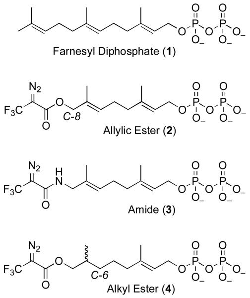
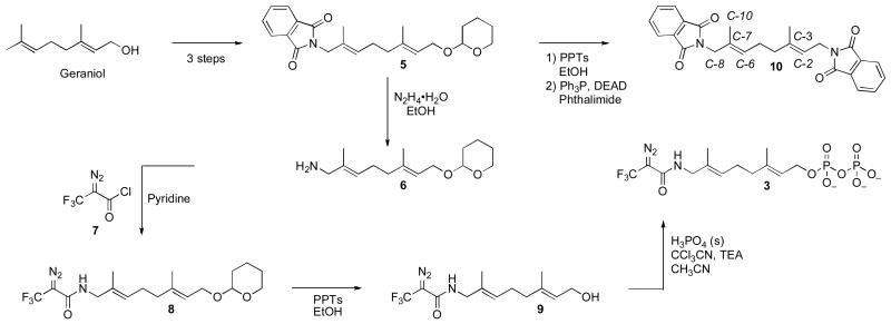
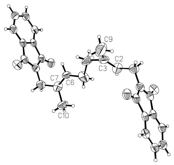
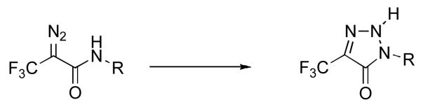
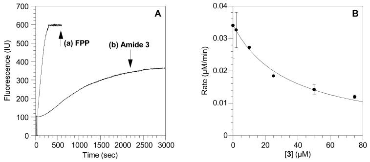
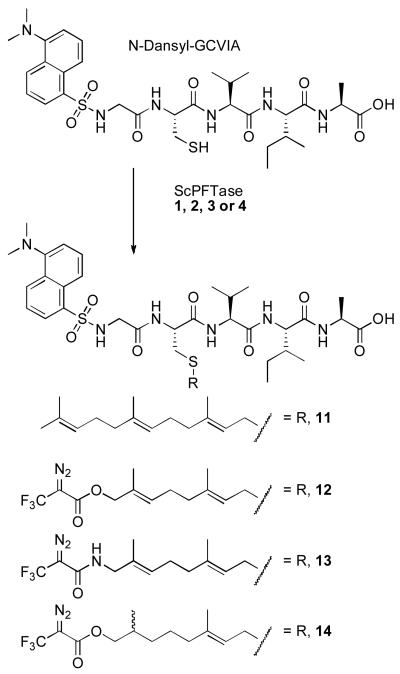
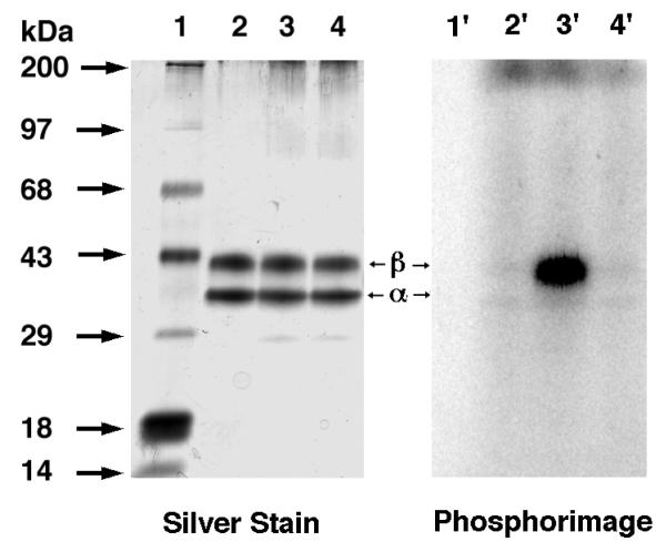
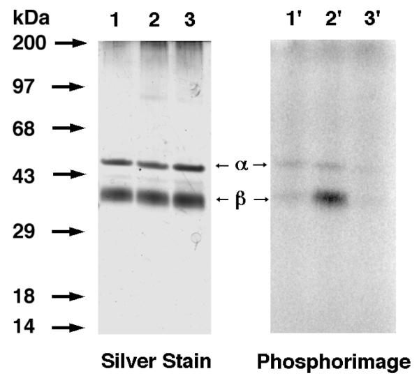
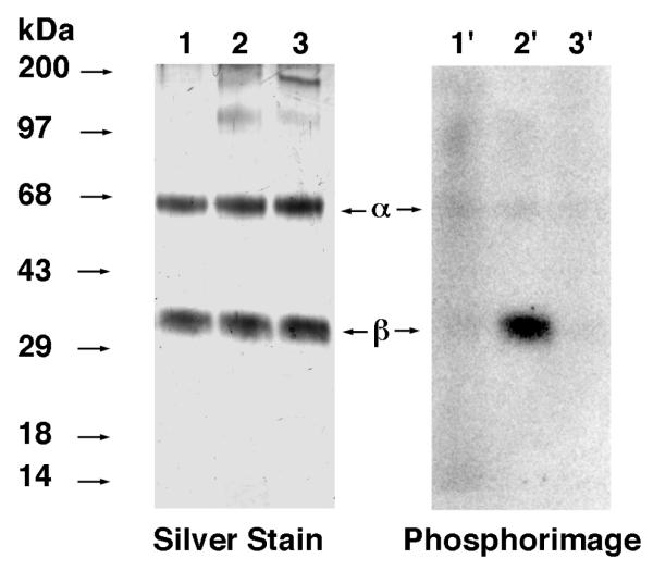
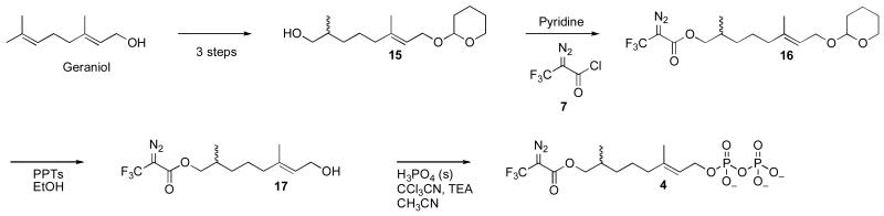
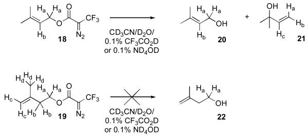
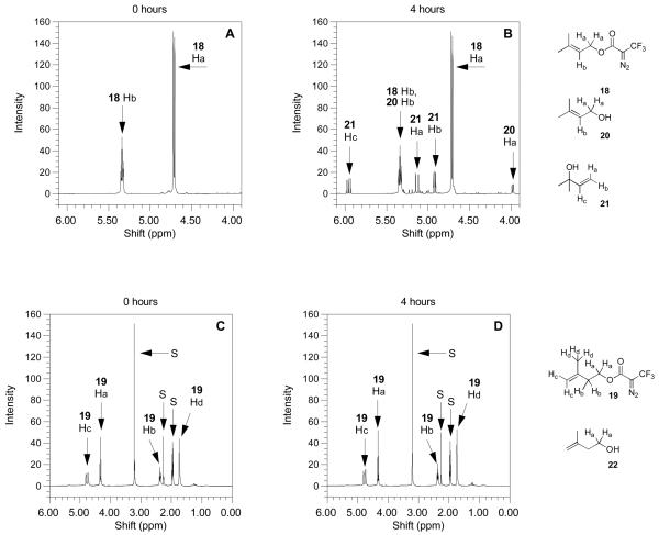
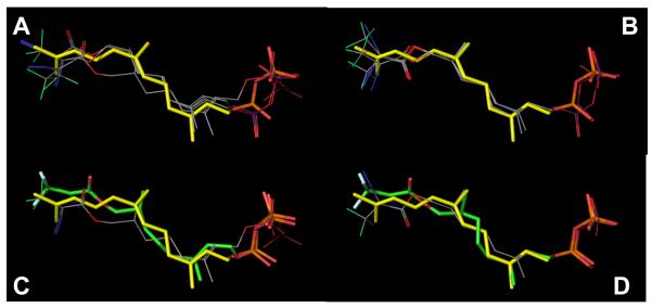
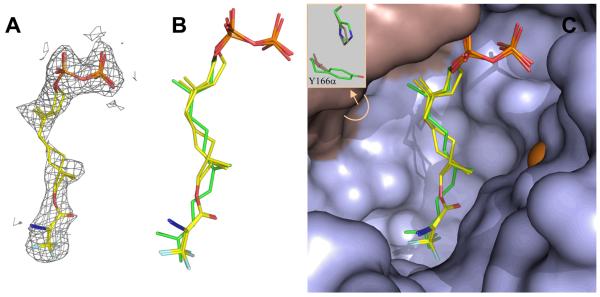
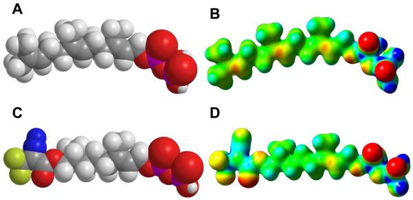
Photoactive analogs of farnesyl diphosphate (FPP) are useful probes in studies of enzymes that employ this molecule as a substrate. Here, we describe the preparation and properties of two new FPP analogs that contain diazotrifluoropropanoyl photophores linked to geranyl diphosphate via amide or ester linkages. The amide-linked analog (3) was synthesized in 32P-labeled form from geraniol in seven steps. Experiments with Saccharomyces cerevisiae protein farnesyltransferase (ScPFTase) showed that 3 is an alternative substrate for the enzyme. Photolysis experiments with [(32)P]3 demonstrate that this compound labels the beta-subunits of both farnesyltransferase and geranylgeranyltransferase (types 1 and 2). However, the amide-linked probe 3 undergoes a rearrangement to a photochemically unreactive isomeric triazolone upon long term storage making it inconvenient to use. To address this stability issue, the ester-linked analog 4 was prepared in six steps from geraniol. Computational analysis and X-ray crystallographic studies suggest that 4 binds to protein farnesyl transferase (PFTase) in a similar fashion as FPP. Compound 4 is also an alternative substrate for PFTase, and a 32P-labeled form selectively photocrosslinks the beta-subunit of ScPFTase as well as E. coli farnesyldiphosphate synthase and a germacrene-producing sesquiterpene synthase from Nostoc sp. strain PCC7120 (a cyanobacterial source). Finally, nearly exclusive labeling of ScPFTase in crude E. coli extract was observed, suggesting that [32P]4 manifests significant selectivity and should hence be useful for identifying novel FPP-utilizing enzymes in crude protein preparations.


Figures

Figure 1
Figure 1
Farnesyl diphosphate and related DATFP-containing photoprobes.
Scheme 1
Scheme 1
Synthesis of amide-linked photoaffinity analog 3.
Figure 2
Figure 2
Structure of compound 10 determined by X-ray crystallography to establish the E stereochemistry of the C-6 alkene.
Figure 3
Figure 3
Rearrangement of DATFP-amides to triazolones.
Figure 4
Figure 4
Evaluation of amide 3 as an alternative substrate and inhibitor of ScPFTase. (A) Reaction between N-dansyl-GCVIA and FPP or amide 3 catalyzed by ScPFTase monitored by fluorescence spectroscopy. (a) Kinetic data using FPP as a substrate. (b) Kinetic data using amide 3 as a substrate. Fluorescence was monitored at 30°C by excitation at 340 nm and emission at 505 nm. (B) .Inhibition of ScPFTase-catalyzed farnesylation of N-dansyl-GCVIA by amide 3. Reactions contained 2.0 μM FPP, 2.0 μM N-dansyl-GCVIA and 3 at varying concentrations and were monitored using a continuous spectrofluorometric assay. Each point is the average of 2-3 determinations with the error bars indicating the standard error for each measurement.
Scheme 2
Scheme 2
Structures of ScPFTase modified N-dansyl-GCVIA peptide products. 11: peptide product from reaction with FPP (1); 12: peptide product from reaction with allylic ester 2; 13: peptide product from reaction with amide 3; 14: peptide product from reaction with alkyl ester 4.
Figure 5
Figure 5
Analysis of photolabeling of ScPFTase with DATFP-amide ([32P]3) by SDS-PAGE. Lanes 1 and 1′: molecular weight standards. Lanes 2 and 2′: sample containing PFTase and [32P]3, no UV irradiation. Lanes 3 and 3′: PFTase irradiated in the presence of [32P]3. Lanes 4 and 4′: PFTase irradiated in the presence of [32P]3 and FPP (substrate). Lanes 1, 2, 3, and 4 show the silver-stained proteins. Lanes 1′, 2′, 3′, and 4′ show the radiolabeled proteins.
Figure 6
Figure 6
Analysis of photolabeling of HsPGGTase I with DATFP-amide ([32P]3) by SDS-PAGE. Lanes 1 and 1′ contain samples of PGGTase I and [32P]3 which were not irradiated. Lanes 2 and 2′ contain samples of HsPGGTase I irradiated at 254 nm in the presence of [32P]3. Lanes 3 and 3′ contain samples of PGGTase I irradiated in the presence of [[32P]3 and FPP (substrate). Lanes 1, 2, and 3 show the silver-stained proteins. Lanes 1′, 2′, and 3′ show the radiolabeled proteins.
Figure 7
Figure 7
Analysis of photolabeling of RnPGGTase II with DATFP-amide ([32P]3) by SDS-PAGE. Lanes 1 and 1′: PGGTase II and [32P]3, no UV irradiation. Lanes 2 and 2′: RnGGPTase II and [32P]3, 1 min UV irradiation. Lanes 3 and 3′: RnPGGTase II, [32P]3, and GGPP, 1 min UV irradiation. Lanes 1, 2, and 3 show the silver-stained proteins. Lanes 1′, 2′, and 3′ show the radiolabeled proteins.
Scheme 3
Scheme 3
Synthesis of dihydroester-linked photoaffinity analog 4.
Scheme 4
Scheme 4
DATFP-containing allylic- and alkyl-ester model compounds and their putitive hydrolysis products.
Figure 8
Figure 8
1H-NMR spectra of DATFP-containing model esters obtained under mild acidolytic conditions. Spectra on the left are the compounds before heating and spectra on the right are the compounds after heating at 100°C for 4 h. (A) Compound 18 treated under acidic conditions (CD3CN/D20 10%/TFA-d 0.1%) before heating. (B) Compound 18 treated under acidic conditions (CD3CN/D20 10%/TFA-d 0.1%) after heating to 100°C for 4 h. The appearance of three new doublets of doublets after heating indicates the presence of 21. (C) Compound 19 treated under acidic conditions (CD3CN/D20 10%/TFA-d 0.1%) before heating. (D) Compound 18 treated under acidic conditions (CD3CN/D20 10%/TFA-d 0.1%) after heating to 100°C for 4 h. Compound 19 shows no visible change in the 1H-NMR spectrum after 4 h. In panels (C) and (D), peaks labeled “S” are from D2O, p-xylene (internal standard for integration) and CD3CN.
Figure 9
Figure 9
Reversed-phase HPLC analysis with fluorescence detection of reactions between N-dansyl-GCVIA and FPP (1) or dihydroester 4 catalyzed by ScPFTase. (A) N-dansyl-GCVIA (2.4 μM) incubated with FPP (1, 10 μM) and ScPFTase (24 nM). Chromatogram 1 (bottom): Reaction before the addition of the enzyme with the peptide eluting at tR=25.7 min. Chromatogram 2 (top): Reaction after 5 min with the prenylated peptide (11) appearing at tR=41.5 min. (B) N-dansyl-GCVIA (2.4 μM) incubated with dihydroester (4, 10 μM) and ScPFTase (24 nM). Chromatogram 1 (bottom): Reaction before the addition of the enzyme with the peptide substrate at tR=25.7 min. Chromatogram 2 (top): Reaction after 23 h with the prenylated peptide (14) appearing at tR=36.0 min and some starting material still present.
Figure 10
Figure 10
Inhibition of ScPFTase-catalyzed farnesylation of N-dansyl-GCVIA by dihydroester 4. Reactions contained 2.0 μM FPP, 2.0 μM N-dansyl-GCVIA and 4 at varying concentrations and were monitored using a continuous spectrofluorometric assay. Each point is the average of 3 determinations with the error bars indicating the standard error for each measurement.
Figure 11
Figure 11
Analysis of photolabeling of purified FPP-utilizing enzymes with dihydroester [32P]4. For all samples, photolysis reactions were fractionated by SDS PAGE followed by phosphorimaging analysis to allow visualization of the radiolabeled proteins. In each case, Lane 1 contains enzyme irradiated in the presence of [32P]4 while Lane 2 contains enzyme irradiated in the presence of [32P]4 and FPP (substrate). Where shown, Lane 3 contains [32P]4 incubated in the presence of protein without irradiation. Panel A: Analysis of photolabeling of purified PFTase. Panel B: Analysis of photolabeling of purified EcFPPSase. Panel C: Analysis of photolabeling of purified NoSTSase.
Figure 12
Figure 12
Selective photolabeling of ScyPFTase in crude E. coli extract using dihydroester 4. Analysis of photolabeling of ScPFTase with [32P]4 by SDS-PAGE. Lanes 1 and 1′: Molecular weight standards. Lanes 2 and 2′: Crude ScPFTase irradiated in the presence of [32P]4. Lanes 3 and 3′: Crude ScPFTase irradiated in the presence of [32P]4 and FPP (substrate). Lanes 4 and 4′: Sample containing crude yPFTase and [32P]4, no UV irradiation. Panel A: Sypro Orange-stained proteins. Panel B: Radiolabeled proteins visualized via phosphorimaging analysis.
Figure 13
Figure 13
Results of docking experiments with dihydroester 4 and RnPFTase. Top Left (A): Top 3 poses for the R enantiomer of 4 (line representations) docked into RnPFTase. The position of FPP (stick representation) bound to RnPFTase determined via crystallography (pdb code: 1JCR) is shown for comparison. Top Right (B): Top 3 poses for the S enantiomer of 4 docked into RnPFTase. Bottom Left (C): Comparison of highest-scoring docked pose for the R enantiomer of 4 (line representation) with the structure of the protein-bound R enantiomer determined via crystallography (stick representation, green carbons) and FPP (stick representation, yellow carbons). Bottom Right (D): Comparison of highest-scoring docked pose for the S enantiomer of 4 (line representation) with the structure of the protein-bound S enantiomer determined via crystallography (stick representation) and FPP (stick representation, yellow carbons). Colors: C (line representations: grey, stick representations: yellow [FPP], green [crystallographic 4), F (line representations: green, stick representations: white), N (blue), O (red), P (line representations: purple, stick representations: orange). As noted in the text, the term “docked” pose used here refers to the docked poses that have been subjected to energy minimization.
Figure 14
Figure 14
Structure of dihydroester 4 bound to RnPFTase determined by X-ray crystallography. (A) Electron density for dihydroester 4 (mesh) contoured at 1.0 σ. The structures of the two enantiomers of 4 (shown in stick representations) are fit within the electron density. (B) Structures of the two enantiomers of 4 superimposed with the structure of FPP (green) bound to RnPFTase. (C) Active site of RnPFTase showing structures of the two enantiomers of 4 superimposed with the structure of FPP (green). The insert shows the difference in conformation for Y166α and H210α between the structures of 4 and FPP. Colors: C (yellow), N (blue), O (red), α-subunit (grey), β-subunit (light blue), Zn (orange).
Figure 15
Figure 15
CPK models and molecular electrostatic potential (MEP) maps calculated for FPP and dihydroester 4. Above: CPK model (A) and MEP (B) for FPP. Below: CPK model (C) and MEP (D) for 4. For MEP maps, potentials were plotted on a surface of constant density (density = 0.02). Electronegative areas are shown in red and electropositive areas are shown in blue.

Similar articles

Cited by

References

    1. Cane DE. Sesquiterpene biosynthesis: Cyclization mechanisms. Comp Nat Prod Chem. 1999;2:155–200.
    1. Davis EM, Croteau R. Cyclization enzymes in the biosynthesis of monoterpenes, sesquiterpenes, and diterpenes. Topics Curr Chem. 2000;209:53–95.
    1. Loomis WD, Croteau R. Biochemistry of terpenoids. Biochem Plants. 1980;4:363–418.
    1. Zhang FL, Casey PJ. Protein prenylation: Molecular mechanisms and functional consequences. Ann Rev Biochem. 1996;65:241–69. - PubMed
    1. Edelstein RL, Distefano MD. Photoaffinity labeling of yeast farnesyl protein transferase and enzymic synthesis of a Ras protein incorporating a photoactive isoprenoid. Biochem Biophys Res Comm. 1997;235:377–82. - PubMed

Publication types

MeSH terms