Review
. 2010 Jan;33(1):1-9.
doi: 10.1016/j.tins.2009.11.002. Epub 2009 Dec 1.

Recovery of consciousness after brain injury: a mesocircuit hypothesis

Affiliations
Review

Recovery of consciousness after brain injury: a mesocircuit hypothesis

Nicholas D Schiff. Trends Neurosci. 2010 Jan.

Abstract

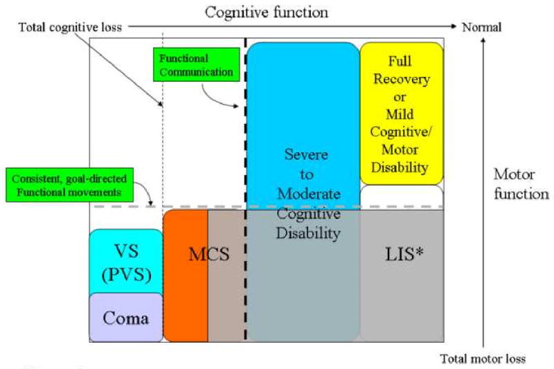
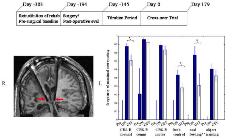
Recovery of consciousness following severe brain injuries can occur over long time intervals. Importantly, evolving cognitive recovery can be strongly dissociated from motor recovery in some individuals, resulting in underestimation of cognitive capacities. Common mechanisms of cerebral dysfunction that arise at the neuronal population level may explain slow functional recoveries from severe brain injuries. This review proposes a "mesocircuit" model that predicts specific roles for different structural and dynamic changes that may occur gradually during recovery. Recent functional neuroimaging studies that operationally identify varying levels of awareness, memory and other higher brain functions in patients with no behavioral evidence of these cognitive capacities are discussed. Measuring evolving changes in underlying brain function and dynamics post-injury and post-treatment frames future investigative work.


Figures

Figure 1
Figure 1. Correspondence of cognitive and motor impairment associated with disorders consciousness arising following severe brain injuries
The distinctions among clinical disorders of consciousness can be best captured on a two-dimensional axis by comparing degree of impaired cognitive function against degree of motor function. At the bottom left of Figure the functional equivalence of coma and vegetative state (VS) as unconscious brain states is indicated by their placement to the left of the vertical dotted line indicating total loss of cognitive function. The large grey box in Figure 1, is placed to mark the high-degree of uncertainty associated with identifying the cognitive capacities of patients with no controllable motor output channel whose clinical bedside examination may range from minimally conscious state (MCS) to locked-in state (LIS), Note the * indicates that the locked-in state is not a disorder of consciousness and LIS patients retain normal cognitive function by definition. Establishing a true cognitive level for many patients who behaviorally cannot reliably signal through controlled goal-directed movements (dashed horizontal line) is possible at present. Such patients may retain varying levels of cognitive processing capabilities, awareness, memory and other higher brain functions without detection. Disentangling the potential for cognitive function in setting of severe limitations of motor control and sensorimotor integration mechanisms is among the most important challenges presented by new understanding of the recovery process following severe brain injuries.
Figure 2
Figure 2. Comparison of regions of central thalamus involved in focal and diffuse injuries producing global impairments of consciousness
A. Focal injury patterns in the central thalamus associated with coma, vegetative state, and minimally conscious state (adapted from [15]). Red circle indicates anterior intralaminar nuclei and surrounding regions), green circle includes area of red circle and more caudal and medial components of the posterior intralaminar region. B. Regional neuronal cell loss in central thalamus following severe traumatic brain injuries indexed by functional outcomes [12]. Moderately disabled patients have cell loss restricted to the anterior intralaminar regions (red circle). Severely disabled patients have neuronal loss in more caudal and medial components of the central thalamus including the medial aspects of the posterior intralaminar nuclei. Permanent VS is associated with broad loss of central thalamic neurons including the large lateral component of the posterior intralaminar group (the centromedian nucleus) [14, 24]. Figure element of thalamic anatomy adapted from [74] with permission.
Figure 3
Figure 3. Proposed “mesocircuit” model underlying forebrain dysfunction and interventions in severe brain injuries
A proposed ‘mesocircuit’ that explains the vulnerability of the anterior forebrain (frontal/prefrontal cortical-striatopallidal thalamocortical loop systems) following multi-focal brain injuries that produce widespread deafferentation or neuronal cell loss. The thalamocortical projections of the central thalamus are proposed to play an important role in observed reduction of cerebral metabolism in this mesocircuit following different mechanisms of brain injury [42, 48]; these projections have a strong activating role strongly driving both cortical and striatal neurons [47, 30,31]. The medium spiny neurons (MSN) of the striatum which send inhibitory projections to the globus pallidus interna require high levels of background synaptic activity and dopaminergic neuromodulation to maintain firing rates [46]. Without MSN output the globus pallidus interna tonically inhibits the central thalamus potentially catalyzing a shut down of the anterior forebrain. Down-regulation of activity within the mesocircuit is predicted to have a broad modulatory impact on the global dynamics of the dominant corticothalamic system [25, 26, 28, 32, 73]; specific changes within the cortico-striatopallidal thalamocortical system identified with alterations of consciousness associated with sleep and anesthesia support this inference [55, 58, 61]. The mesocircuit model also economically accounts for the mix of interventions that have been noted in some patients to restore functions associated with these forebrain systems (e.g. dopaminergic agents, zolpidem, and electrical brain stimulation; see text for further discussion).
Figure 4
Figure 4. Changes in cerebral metabolism associated with zolpidem administration in severe brain-injury
Fluorodeoxyglucose positron emission tomography studies by Brefel-Courbon et al. [42] of a severe brain injured patient in minimally conscious state before and after administration of the sedative agent zolpidem (‘Ambien’). In the off drug state (top panels) marked anterior forebrain hypometabolism is noted bilaterally in frontal/prefrontal cortex, thalami, and striatum. Following zolpidem administration broad increases of metabolic rates are observed in these regions.
Figure 5
Figure 5. Central thalamic DBS in the minimally conscious state
A. Timeline of single-subject study of deep brain stimulation in the central thalamus in a patient remaining in MCS for 6 years B. Location of electrode lead placements within central thalamus of patient's right (R) and left (L) hemispheres displayed in T1 weighted MRI coronal image and marked with red arrows. C. Comparison of pre-surgical baselines and DBS ON and DBS OFF periods during a six month cross-over trial. Behavioral baseline evaluations measured using a standardized quantitative assessment tool, the Coma Recovery Scale Revised (CRS-R) were obtained 4 months prior to surgery and for 2 months following surgery prior to titration testing of stimulation parameters [see Ref #64 for details] showed no change in behavioral responsiveness as compared to functional levels measured more than 2 years before the start of the trial. During the early titration testing of electrical stimulation the patient demonstrated immediate and accumulating effects of DBS that included the emergence of consistent and intelligible spoken language, recovery of limb control and the capacity for oral feeding (see [64] for details). Following a 5 month titration period of testing combinations of stimulation parameters the patient entered into a blinded six-month, 30 day alternating ON versus OFF (cross-over) study that demonstrated a robust overall effect on behavioral responsiveness measured by CRS-R subscales and supplementary behavioral rating scales. Significant ON versus OFF improvements with electrical were demonstrated for attentive behavior, oral feeding and limb control. All functional testing showed significant improvements when compared against the 6 month pre-stimulation baselines. Importantly, observed carryover effects of improvements from the ON to the OFF state were also identified as seen by the high frequency of OFF stimulation ratings compared to pre-stimulation baselines across all measurements. These effects are comparable to evidence of accumulating behavioral effects of central thalamic electrical stimulation as shown in a rodent model by Herrera and colleagues [66]. Figure elements adapted from Schiff et al [64] with permission.

Similar articles

Cited by

References

    1. Lammi MH, Smith VH, Tate RL, Taylor CM. The minimally conscious state and recovery potential: a follow-up study 2 to 5 years after traumatic brain injury. Arch Phys Med Rehabil. 2005;86(4):746–54. - PubMed
    1. Voss HU, et al. Possible axonal regrowth in late recovery from minimally conscious state. Journal of Clinical Investigation. 2006;116:2005–2011. - PMC - PubMed
    1. Burruss JW, Chacko RC. Episodically remitting akinetic mutism following subarachnoid hemorrhage. J Neuropsychiatry Clin Neurosci 1999. 1999;11(1):100–2. - PubMed
    1. McMillan TM, Herbert CM. Further recovery in a potential treatment withdrawal case 10 years after brain injury. Brain Inj. 2004;18(9):935–40. - PubMed
    1. Macniven JA, Poz R, Bainbridge K, Gracey F, Wilson BA. Emotional adjustment following cognitive recovery from ‘persistent vegetative state’: psychological and personal perspectives. Brain Inj. 2003;17(6):525–33. - PubMed

Publication types

MeSH terms