. 2011 Oct;32(10):1818-26.
doi: 10.1016/j.neurobiolaging.2009.10.017. Epub 2009 Dec 1.

RACK1 is involved in β-amyloid impairment of muscarinic regulation of GABAergic transmission

Affiliations

RACK1 is involved in β-amyloid impairment of muscarinic regulation of GABAergic transmission

Wenhua Liu et al. Neurobiol Aging. 2011 Oct.

Abstract

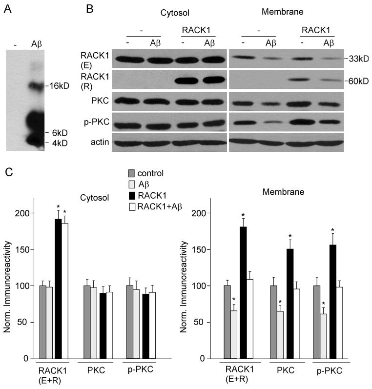
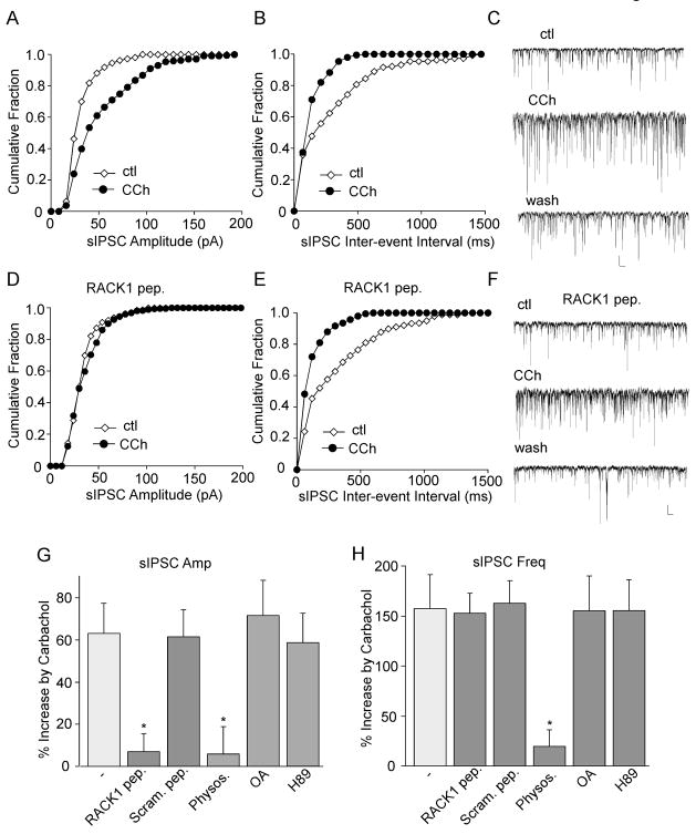
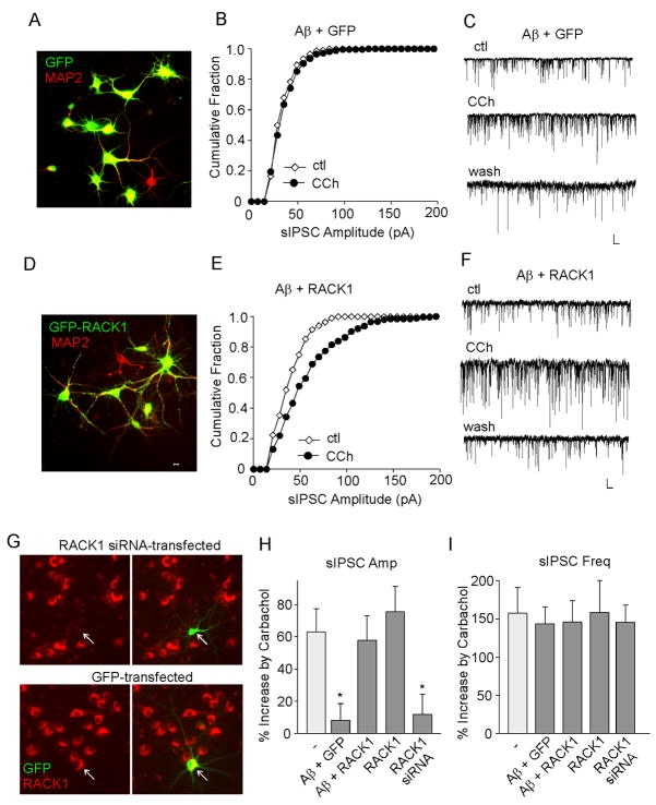
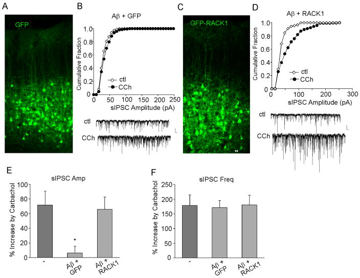
RACK1 (receptor for activated C-kinase 1), an anchoring protein that shuttles activated PKC to cellular membranes, plays an important role in PKC-mediated signal transduction pathways. A significant loss of RACK1 has been found in the brain of aging animals and Alzheimer's disease (AD) patients, which implicates the potential involvement of RACK1 in altered PKC activation associated with dementia. Our previous studies have demonstrated that GABAergic inhibition in prefrontal cortex, which is important for cognitive processes like "working memory", is regulated by muscarinic receptors via a PKC-dependent mechanism, and this effect is impaired by β-amyloid peptide (Aβ). In this study, we found that Aβ oligomers decreased RACK1 distribution in the membrane fraction of cortical neurons. Moreover, overexpression of RACK1 rescued the effect of muscarinic receptors on GABAergic transmission in Aβ-treated cortical cultures in vitro and Aβ-injected cortical neurons in vivo. These results suggest that the Aβ-induced loss of RACK1 distribution in the cell membrane may underlie the Aβ impairment of muscarinic regulation of PKC and GABAergic transmission. Thus, RACK1 provides a potential therapeutic target that can restore some of the impaired cellular processes by Aβ.


Figures

FIG. 1
FIG. 1
42 treatment decreases RACK1, total and activated PKC in membrane fractions of cultured cortical neurons. A, Western blots showing the oligomeric Aβ B, Western blots showing endogenous (E) and recombinant (R) RACK1, PKC, p-PKC and actin in cytosolic and membrane fractions from cortical cultures (uninfected or infected with GFP-RACK1 virus) treated without or with Aβ oligomer (1 μM, 48 hrs). C, Quantifications by densitometric analysis of RACK1 (E+R), PKC, p-PKC in cytosolic and membrane fractions from non-treated or Aβ-treated cortical cultures without or with RACK1 overexpression. Protein levels (normalized to actin) were expressed as the percent of controls. *: p < 0.01, ANOVA, compared to untreated and uninfected neurons (control).
FIG. 2
FIG. 2
RACK1 is involved in muscarinic regulation of sIPSC amplitude. A–C, Cumulative plots of the distribution of sIPSC amplitudes (A) and inter-event interval (B), and representative sIPSC traces (C) in a cultured cortical neuron before (ctl) and after carbachol (CCh, 20 μM) application. D–F, Cumulative plots of the distribution of sIPSC amplitudes (D) and inter-event interval (E), and representative sIPSC traces (F) showing the effect of carbachol in a cultured cortical neuron dialyzed with a RACK1 peptide (40 μM). Scale bars: 30 pA, 1s. G,H, Cumulative data (mean ± SEM) showing the percent increase of sIPSC amplitude (G) or frequency (H) by carbachol in the absence or presence of RACK1 peptide, a scrambled control peptide (40 μM), acetylcholinesterase (AChE) inhibitor physostigmine (40 μM), protein phosphatase inhibitor okadaic acid (OA, 1 μM), or PKA inhibitor H89 (10 μM). *: p < 0.01, ANOVA, compared to the effect of carbachol in the control condition (−).
FIG. 3
FIG. 3
Overexpression of RACK1 restores the Aβ-induced loss of muscarinic regulation of sIPSC amplitudes in cortical cultures. A,D, Immunocytochemical images of MAP2(red)-stained cortical cultures infected with GFP (A) or GFP-RACK1 (D) Sindbis viruses. B,E, Cumulative plots of the distribution of sIPSC amplitudes showing the effect of carbachol (CCh, 20 μM) in A (1 μM, 2–3 days)-treated cortical cultures infected with GFP (B) or GFP-RACK1 (E) Sindbis viruses. C,F, Representative sIPSC traces from the neurons used to construct B and E. Scale bars: 30 pA, 1s. G, Immunostaining of RACK1 (red) in cultured cortical neurons transfected with RACK1 siRNA (co-transfected with GFP) or GFP alone. Arrowheads point to GFP+ neurons. H, I, Cumulative data (mean ± SEM) showing the percent increase of sIPSC amplitude (H) or frequency (I) by carbachol in cortical neurons under different conditions. *: p < 0.01, ANOVA, compared to the effect of carbachol in the control condition (−).
FIG. 4
FIG. 4
RACK1 expression restores the Aβ-induced loss of muscarinic regulation of sIPSC amplitudes in vivo. A,C, Immunocytochemical images of cortical slices from rats stereotaxically injected with GFP (A) or GFP-RACK1 (C) Sindbis viruses. B,D, Cumulative plots of the distribution of sIPSC amplitudes showing the effect of carbachol (CCh, 20 μM) in cortical neurons from A (1 μM)-injected rats that were infected with GFP (B) or GFP-RACK1 (D) Sindbis viruses. Inset: representative sIPSC traces. Scale bars: 30 pA, 1s. E,F, Cumulative data (mean ± SEM) showing the percent increase of sIPSC amplitude (E) or frequency (F) by carbachol in cortical neurons (from Aβ-injected rats) overexpressing GFP or GFP-RACK1. *: p < 0.01, ANOVA, compared to the effect of carbachol in the control condition (−).

Similar articles

Cited by

References

    1. Araujo DM, Lapchak PA, Robitaille Y, Gauthier S, Quirion R. Differential alteration of various cholinergic markers in cortical and subcortical regions of human brain in Alzheimer’s disease. J Neurochem. 1988;50(6):1914–1923. - PubMed
    1. Battaini F, Pascale A, Paoletti R, Govoni S. The role of anchoring protein RACK1 in PKC activation in the ageing rat brain. Trends Neurosci. 1997;20:410–415. - PubMed
    1. Battaini F, Pascale A, Lucchi L, Pasinetti GM, Govoni S. Protein kinase C anchoring deficit in postmortem brains of Alzheimer’s disease patients. Exp Neurol. 1999;159:559–564. - PubMed
    1. Behn-Krappa A, Newton AC. The hydrophobic phosphorylation motif of conventional protein kinase C is regulated by autophosphorylation. Curr Biol. 1999;9:728–37. - PubMed
    1. Broersen LM, Heinsbroek RPW, de Bruin JPC, Uylings HBM, Oliver B. The role of the medial prefrontal cortex of rats in short term memory functioning: further support for involvement of cholinergic, rather than dopaminergic mechanisms. Brain Res. 1995;674:221–229. - PubMed

Publication types

MeSH terms