. 2010 Feb;9(2):327-35.
doi: 10.1074/mcp.M900248-MCP200. Epub 2009 Nov 27.

Sensitive plasma protein analysis by microparticle-based proximity ligation assays

Affiliations

Sensitive plasma protein analysis by microparticle-based proximity ligation assays

Spyros Darmanis et al. Mol Cell Proteomics. 2010 Feb.

Abstract

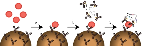
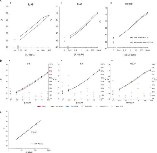
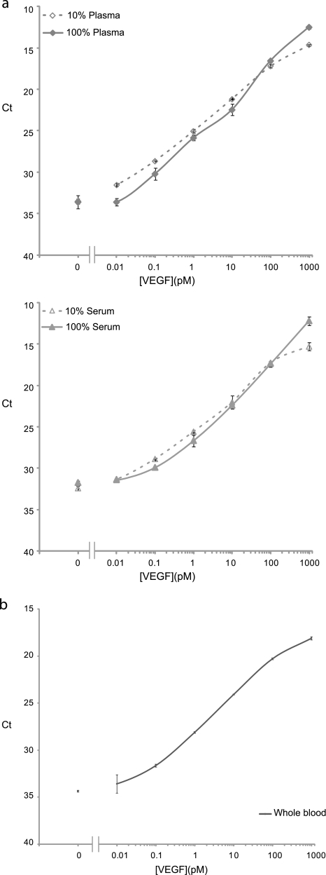
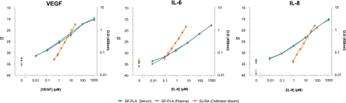
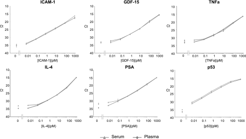
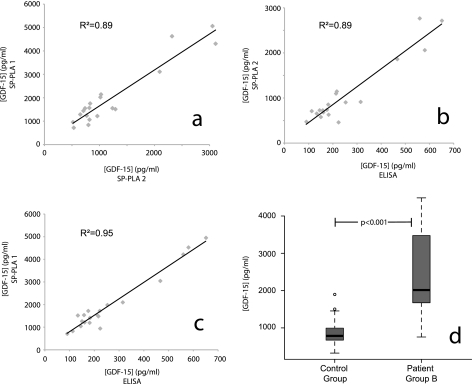
Detection of proteins released in the bloodstream from tissues damaged by disease can promote early detection of pathological conditions, differential diagnostics, and follow-up of therapy. Despite these prospects and a plethora of candidate biomarkers, efforts in recent years to establish new protein diagnostic assays have met with limited success. One important limiting factor has been the challenge of detecting proteins present at trace levels in complex bodily fluids. To achieve robust, sensitive, and specific detection, we have developed a microparticle-based solid-phase proximity ligation assay, dependent on simultaneous recognition of target proteins by three antibody molecules for added specificity. After capture on a microparticle, solid-phase pairs of proximity probes are added followed by washes, enabling detection and identification of rare protein molecules in blood while consuming small amounts of sample. We demonstrate that single polyclonal antibody preparations raised against target proteins of interest can be readily used to establish assays where detection depends on target recognition by three individual antibody molecules, recognizing separate epitopes. The assay was compared with state-of-the-art sandwich ELISAs for detection of vascular endothelial growth factor, interleukin-8 and interleukin-6, and it was found to be superior both with regard to dynamic range and minimal numbers of molecules detected. Furthermore, the assays exhibited excellent performance in undiluted plasma and serum as well as in whole blood, producing comparable results for nine different antigens. We thus show that solid-phase proximity ligation assay is suitable for validation of a variety of protein biomarkers over broad dynamic ranges in clinical samples.


Figures

Fig. 1.
Fig. 1.
Schematic description of SP-PLA method. A, samples are incubated with antibodies preimmobilized on microparticles. B, next, microparticles are washed and incubated with pairs of PLA probes. C, finally, oligonucleotides on PLA probes are ligated upon proximal binding of a common antigen and addition of a connector oligonucleotide. This is followed by amplification and detection of the ligated products by quantitative real time PCR, the primers of which are indicated by arrows.
Fig. 2.
Fig. 2.
Performance of microparticle-based SP-PLA and comparison with tube-based assay. a, comparison of microparticle-based (squares) and tube-based assay (diamonds) for detection of IL-6 (I), IL-8 (II), and VEGF (III) in buffer. All measurements were performed at least in triplicates. Error bars indicate SD. b, SP-PLA performance for detection of IL-6 (I), IL-8 (II), and VEGF (III) in buffer (squares), 10% serum (triangles), and 10% plasma (diamonds) with corresponding CV% values for every data point for buffer (open squares), 10% serum (open triangles), and 10% plasma (open diamonds). c, 2-fold dilutions of IL-8 in 10% plasma to estimate precision. The y axes display threshold cycle values of real time PCR assays; the x axes display concentrations of the investigated proteins in pm.
Fig. 3.
Fig. 3.
Performance of SP-PLA in complex biological samples. a, comparison of performance of SP-PLA in 100% serum (open triangles) and plasma (open diamonds) and 10% serum (triangles) and plasma (diamonds) for the detection of VEGF. b, detection of VEGF by SP-PLA in whole blood. Error bars indicate SD.
Fig. 4.
Fig. 4.
Comparison of SP-PLA with sandwich ELISA for detection of VEGF, IL-6, and IL-8. For SP-PLA, the proteins were spiked in 10% serum (triangles) and 10% plasma (diamonds), and for ELISA, the proteins were spiked in the 10% calibrator diluent for serum and plasma provided by the manufacturer (circles). The primary y axes display threshold cycle values of real time PCR for SP-PLA; the secondary y axes display OD measured at 450 nm for ELISA. x axes display concentration of the analyzed proteins in the 10-fold dilutions of each protein in the 50 μl SP-PLA reactions and the 200 μl ELISAs. All measurements were performed at least in triplicates. Error bars indicate SD.
Fig. 5.
Fig. 5.
Detection of ICAM-1, GDF-15, TNFα, IL-4, PSA, and p53 with SP-PLA in 10% serum (triangles) and 10% plasma (diamonds). y axes display threshold cycle values of real time PCR assays; x axes display concentration of each antigen in pm.
Fig. 6.
Fig. 6.
Measurements of GDF-15 in patient samples as analyzed in two independent SP-PLA tests and in one sandwich ELISA. a, GDF-15 was measured by SP-PLA in 20 human plasma samples obtained from patient group A on two separate occasions (SP-PLA1 and SP-PLA2). Values of GDF-15 in pg/ml obtained in assay 1 are plotted on the y axis, whereas values obtained from assay 2 are plotted on the x axis. b and c, scatter plots showing the correlation between ELISA and each of the two SP-PLA tests (SP-PLA1 and SP-PLA2) for data from the same 20 human plasma samples. Values of GDF-15 in pg/ml obtained from SP-PLA are plotted on the y axis, whereas ELISA values are plotted on the x axis. d, box plots showing differences in levels of GDF-15 in patients belonging to patient group B and in healthy controls. The p value was calculated using a two-sample Wilcoxon rank sum test.

Similar articles

Cited by

References

    1. Anderson N. L., Anderson N. G. ( 2002) The human plasma proteome: history, character, and diagnostic prospects. Mol. Cell. Proteomics 1, 845– 867 - PubMed
    1. Fredriksson S., Gullberg M., Jarvius J., Olsson C., Pietras K., Gústafsdóttir S. M., Ostman A., Landegren U. ( 2002) Protein detection using proximity-dependent DNA ligation assays. Nat. Biotechnol 20, 473– 477 - PubMed
    1. Gustafsdottir S. M., Nordengrahn A., Fredriksson S., Wallgren P., Rivera E., Schallmeiner E., Merza M., Landegren U. ( 2006) Detection of individual microbial pathogens by proximity ligation. Clin. Chem 52, 1152– 1160 - PubMed
    1. Söderberg O., Gullberg M., Jarvius M., Ridderstråle K., Leuchowius K. J., Jarvius J., Wester K., Hydbring P., Bahram F., Larsson L. G., Landegren U. ( 2006) Direct observation of individual endogenous protein complexes in situ by proximity ligation. Nat. Methods 3, 995– 1000 - PubMed
    1. Gustafsdottir S. M., Schlingemann J., Rada-Iglesias A., Schallmeiner E., Kamali-Moghaddam M., Wadelius C., Landegren U. ( 2007) In vitro analysis of DNA-protein interactions by proximity ligation. Proc. Natl. Acad. Sci. U.S.A 104, 3067– 3072 - PMC - PubMed

Publication types