. 2010 Jan 29;285(5):3360-70.
doi: 10.1074/jbc.M109.068650. Epub 2009 Dec 2.

Functions of the alpha, beta, and gamma subunits of UDP-GlcNAc:lysosomal enzyme N-acetylglucosamine-1-phosphotransferase

Affiliations

Functions of the alpha, beta, and gamma subunits of UDP-GlcNAc:lysosomal enzyme N-acetylglucosamine-1-phosphotransferase

Yi Qian et al. J Biol Chem. .

Abstract

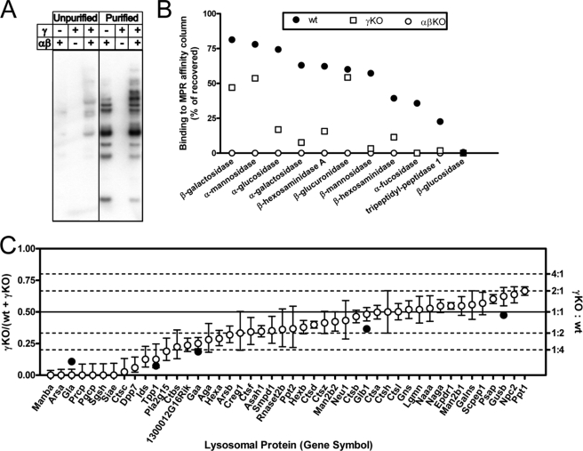
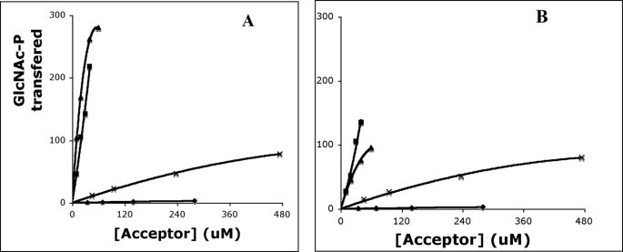
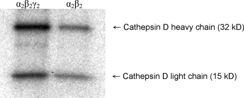
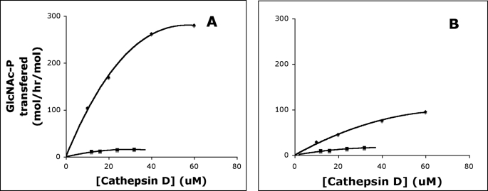
UDP-GlcNAc:lysosomal enzyme N-acetylglucosamine-1-phosphotransferase is an alpha(2)beta(2)gamma(2) hexamer that mediates the first step in the synthesis of the mannose 6-phosphate recognition marker on lysosomal acid hydrolases. Using a multifaceted approach, including analysis of acid hydrolase phosphorylation in mice and fibroblasts lacking the gamma subunit along with kinetic studies of recombinant alpha(2)beta(2)gamma(2) and alpha(2)beta(2) forms of the transferase, we have explored the function of the alpha/beta and gamma subunits. The findings demonstrate that the alpha/beta subunits recognize the protein determinant of acid hydrolases in addition to mediating the catalytic function of the transferase. In mouse brain, the alpha/beta subunits phosphorylate about one-third of the acid hydrolases at close to wild-type levels but require the gamma subunit for optimal phosphorylation of the rest of the acid hydrolases. In addition to enhancing the activity of the alpha/beta subunits toward a subset of the acid hydrolases, the gamma subunit facilitates the addition of the second GlcNAc-P to high mannose oligosaccharides of these substrates. We postulate that the mannose 6-phosphate receptor homology domain of the gamma subunit binds and presents the high mannose glycans of the acceptor to the alpha/beta catalytic site in a favorable manner.


Figures

FIGURE 1.
FIGURE 1.
Analysis of acid hydrolase phosphorylation in wild-type and phosphotransferase-deficient mouse brain. A, Man-6-P-containing glycoproteins in unpurified brain homogenates or in affinity column Man-6-P eluates. B, analysis of binding to CI-MPR column using enzyme activity assays. C, comparison of relative levels of Man-6-P-containing lysosomal proteins in wild-type and γ-knock out samples. Open circles and error bars represent the point estimate and 95% confidence intervals for the proportion of each indicated gene product determined by LC-MS/MS analysis and spectral counting of the affinity-purified samples. Only proteins of interest with ≥30 spectral counts that have been classified as lysosomal Man-6-P-containing glycoproteins or likely candidates are shown (35). Data for all proteins identified and statistical analyses are presented in supplemental Table 1. Filled circles represent the relative proportion (% bound for γ-KO/(% bound for wild type + % bound for γ-KO)) for select enzymes in B that can be attributed to a given gene product. Note that equivalent amounts of affinity-purified samples were used for spectral count analysis, yet the yield of protein eluted from the column was ∼2-fold greater for the wild-type compared with the γ-knock-out sample. Thus, the spectral count analysis overestimates (or represents an upper limit for) the relative amount of a given protein in the γ-knock-out sample.
FIGURE 2.
FIGURE 2.
GlcNAc-1-phosphotransferase activity toward various acceptors. Cathepsin D (▴), NPC2 (■), RNase B (×), and soybean agglutinin (♦) were compared as acceptors for α2β2γ2 (A) and α2β2 forms (B) of GlcNAc-1-phosphotransferase. Various concentrations of the substrates and either 1.2 μg of the α2β2γ2 or 0.97 μg of the α2β2 GlcNAc-1-phosphotransferase were incubated in 50 mm Tris-HCl, pH 7.4, 10 mm MgCl2, 10 mm MnCl2, 75 μm UDP-[3H]GlcNAc (1 μCi), 2 mg/ml bovine serum albumin in a final volume of 50 μl for 1 h at 37 °C. The incorporated [3H]GlcNAc-P was determined by scintillation counting. The activity of the GlcNAc-1-phosphotransferase was expressed as moles of [3H]GlcNAc-P transferred per h/mol of the GlcNAc-1-phosphotransferase.
FIGURE 3.
FIGURE 3.
Phosphorylation of individual glycosylation sites of cathepsin D. α2β2γ2 (left) and α2β2 forms (right) of GlcNAc-1-phosphotransferase (2.5 μg of each) were incubated with 5 μm cathepsin D in the presence of 75 μm [β-32P]UDP-GlcNAc (10 μCi) in a volume of 50 μl. After 30 min, the reaction mixtures were diluted to 500 μl of Tris-buffered saline, 1% Triton X-100 followed by immunoprecipitation with anti-cathepsin D antibody, SDS-PAGE, and autoradiography. The upper band represents the cathepsin D heavy chain, and the lower band represents the light chain.
FIGURE 4.
FIGURE 4.
Effect of lysine modification on cathepsin D phosphorylation. α2β2γ2 (A) and α2β2 forms (B) of GlcNAc-1-phosphotransferase were incubated with increasing concentrations of cathepsin D (♦) and SNA-treated cathepsin D (■) in 50 mm Tris-HCl, pH 7.4, 10 mm MgCl2, 10 mm MnCl2, 75 μm UDP-[3H]GlcNAc (1 μCi), 2 mg/ml bovine serum albumin in a final volume of 50 μl for 1 h at 37 °C. The incorporated [3H]GlcNAc-P was determined by scintillation counting. The activity of the GlcNAc-1-phosphotransferase was expressed as moles of [3H]GlcNAc-P transferred per h/mol of the GlcNAc-1-phosphotransferase.

References

    1. Braulke T., Bonifacino J. S. (2009) Biochim. Biophys. Acta 1793, 605–614 - PubMed
    1. Bao M., Booth J. L., Elmendorf B. J., Canfield W. M. (1996) J. Biol. Chem. 271, 31437–31445 - PubMed
    1. Raas-Rothschild A., Cormier-Daire V., Bao M., Genin E., Salomon R., Brewer K., Zeigler M., Mandel H., Toth S., Roe B., Munnich A., Canfield W. M. (2000) J. Clin. Invest. 105, 673–681 - PMC - PubMed
    1. Tiede S., Storch S., Lübke T., Henrissat B., Bargal R., Raas-Rothschild A., Braulke T. (2005) Nat. Med. 11, 1109–1112 - PubMed
    1. Kudo M., Bao M., D'Souza A., Ying F., Pan H., Roe B. A., Canfield W. M. (2005) J. Biol. Chem. 280, 36141–36149 - PubMed

Publication types

Substances