. 2010 Jan 22;285(4):2333-9.
doi: 10.1074/jbc.M109.090522. Epub 2009 Dec 2.

Transforming growth factor beta-activated kinase 1 (TAK1) kinase adaptor, TAK1-binding protein 2, plays dual roles in TAK1 signaling by recruiting both an activator and an inhibitor of TAK1 kinase in tumor necrosis factor signaling pathway

Affiliations

Transforming growth factor beta-activated kinase 1 (TAK1) kinase adaptor, TAK1-binding protein 2, plays dual roles in TAK1 signaling by recruiting both an activator and an inhibitor of TAK1 kinase in tumor necrosis factor signaling pathway

Peter Broglie et al. J Biol Chem. .

Abstract

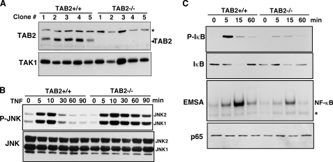
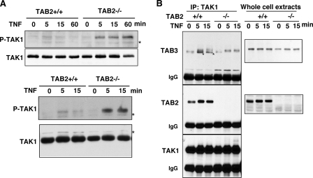
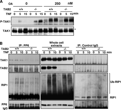
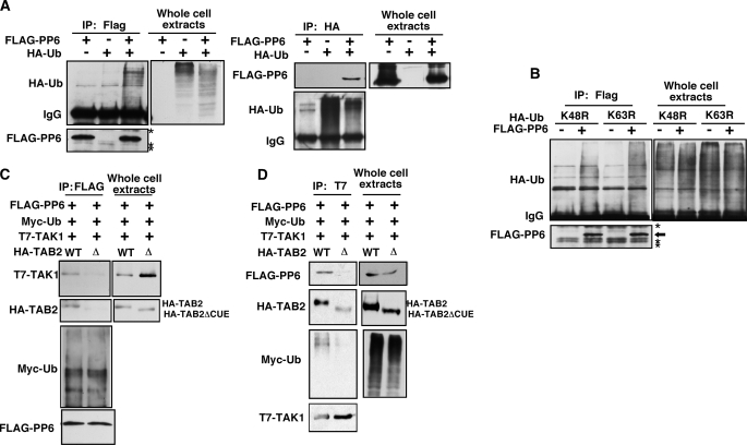
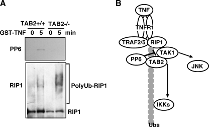
Transforming growth factor beta-activated kinase 1 (TAK1) kinase is an indispensable signaling intermediate in tumor necrosis factor (TNF), interleukin 1, and Toll-like receptor signaling pathways. TAK1-binding protein 2 (TAB2) and its closely related protein, TAB3, are binding partners of TAK1 and have previously been identified as adaptors of TAK1 that recruit TAK1 to a TNF receptor signaling complex. TAB2 and TAB3 redundantly mediate activation of TAK1. In this study, we investigated the role of TAB2 by analyzing fibroblasts having targeted deletion of the tab2 gene. In TAB2-deficient fibroblasts, TAK1 was associated with TAB3 and was activated following TNF stimulation. However, TAB2-deficient fibroblasts displayed a significantly prolonged activation of TAK1 compared with wild type control cells. This suggests that TAB2 mediates deactivation of TAK1. We found that a TAK1-negative regulator, protein phosphatase 6 (PP6), was recruited to the TAK1 complex in wild type but not in TAB2-deficient fibroblasts. Furthermore, we demonstrated that both PP6 and TAB2 interacted with the polyubiquitin chains and this interaction mediated the assembly with TAK1. Our results indicate that TAB2 not only activates TAK1 but also plays an essential role in the deactivation of TAK1 by recruiting PP6 through a polyubiquitin chain-dependent mechanism.


Figures

FIGURE 1.
FIGURE 1.
MAPK and NF-κB pathways are activated in response to TNF independently of TAB2. A, map3k7ip3flox/flox dermis fibroblasts were infected with a control or a Cre-expressing retrovirus. More than 20 independent control or Cre-expressing clones were isolated. The expression levels of TAB2 were confirmed by immunoblot analysis using an anti-TAB2 antibody. Representative immunoblots are shown. Anti-TAK1 immunoblotting was used as a loading control. Asterisk indicates a nonspecific band. B, control (TAB2+/+) and TAB2-deficient (TAB2−/−) fibroblasts were stimulated by TNF (20 ng/ml), and JNK activation was detected by immunoblots with anti-phospho-JNK (upper panel) and anti-JNK (lower panel). C, TAB2+/+ and TAB2−/− fibroblasts were stimulated with TNF (20 ng/ml), and activation of NF-κB was detected by immunoblots with anti-phospho-IκB (top panel) and anti-IκB (second panel) and an NF-κB electrophoresis mobility shift assay (third panel). Total p65 NF-κB levels were determined by anti-p65 immunoblots. Asterisk indicates a nonspecific band.
FIGURE 2.
FIGURE 2.
TNF-induced TAK1 activation is sustained in TAB2-deficient fibroblasts. A, two independent clones of control (TAB2+/+) and TAB2-deficient (TAB2−/−) fibroblasts were stimulated with TNF (20 ng/ml). An activated form of TAK1 was detected by anti-phospho-TAK1 (Thr-187) immunoblotting, and the total amount of TAK1 was determined using an anti-TAK1 antibody. Data are representative of six different clones. Asterisks indicate nonspecific bands. B, TAB2+/+ and TAB2−/− fibroblasts were stimulated with TNF (20 ng/ml), and proteins from cell lysates were immunoprecipitated (IP) with anti-TAK1 antibody. TAB2 and TAB3 co-precipitation was analyzed using anti-TAB2 and anti-TAB3 antibodies. Anti-TAB3 was weakly cross-reacted with TAB2. The bands indicated with asterisks are TAB2 that were reacted with anti-TAB3.
FIGURE 3.
FIGURE 3.
TAB2 is essential for TAK1-PP6 interaction. A, control (TAB2+/+) and TAB2-deficient (TAB2−/−) fibroblasts were pretreated with 250 nm OA for 4 h before TNF treatment (20 ng/ml). Activation of TAK1 was detected by using anti-phospho-TAK1 (Thr-187) antibodies, and the total amounts of TAK1 were determined using an anti-TAK1 antibody. Asterisks indicate nonspecific bands. B, TAB2+/+ and TAB2−/− fibroblasts were stimulated with TNF (20 ng/ml), and proteins from cell lysates were immunoprecipitated (IP) with an anti-PP6 antibody or control IgG. TAK1, TAB2, and RIP1 co-precipitation was determined using anti-TAK1, anti-TAB2, and anti-RIP1 antibodies.
FIGURE 4.
FIGURE 4.
Interaction between TAK1 and PP6 is polyubiquitin-dependent. A, 293 cells were transfected with expression vectors for FLAG-tagged PP6 (FLAG-PP6) and HA-tagged ubiquitin (HA-Ub). Cells were lysed, and proteins were immunoprecipitated (IP) with anti-FLAG antibody (left panels) or anti-HA antibody (right panels). Immunoprecipitates and whole cell lysates were analyzed by immunoblotting with an anti-HA antibody and anti-FLAG. Asterisks indicate nonspecific bands. B, 293 cells were transfected with expression vectors for FLAG-tagged PP6 (FLAG-PP6) and HA-tagged K48R or K63R mutant ubiquitin. Cells were lysed, and proteins were immunoprecipitated with anti-FLAG antibody. Immunoprecipitates and whole cell lysates were analyzed by immunoblotting with an anti-HA antibody and anti-FLAG. Asterisks indicate nonspecific bands. C, 293 cells were transfected with expression vectors for T7-tagged TAK1 (T7-TAK1), FLAG-tagged PP6 (FLAG-PP6), Myc-tagged ubiquitin (Myc-Ub), and HA-tagged TAB2 (HA-TAB2) wild-type (WT) or the CUE domain-lacking mutant (Δ). Cells were lysed, and FLAG-PP6 was immunoprecipitated with anti-FLAG antibody. Immunoprecipitates and whole cell lysates were analyzed by immunoblotting with anti-T7, anti-HA, anti-Myc, and anti-FLAG antibodies. D, cell lysates were immunoprecipitated with anti-T7 antibody. The immunoprecipitates and whole cell lysates were analyzed with anti-FLAG, anti-HA, anti-Myc, and anti-T7 antibodies.
FIGURE 5.
FIGURE 5.
PP6 is recruited to TNF receptor complex. A, control (TAB2+/+) and TAB2-deficient (TAB2−/−) fibroblasts were left untreated or stimulated with GST-TNF (1 μg/ml) for 5 min. GST pulldown was performed, and the precipitates were analyzed with anti-PP6 and anti-RIP1 antibodies. B, model.

Similar articles

Cited by

References

    1. Kawai T., Akira S. (2007) Trends Mol. Med. 13, 460–469 - PubMed
    1. Ninomiya-Tsuji J., Kishimoto K., Hiyama A., Inoue J., Cao Z., Matsumoto K. (1999) Nature 398, 252–256 - PubMed
    1. Sato S., Sanjo H., Takeda K., Ninomiya-Tsuji J., Yamamoto M., Kawai T., Matsumoto K., Takeuchi O., Akira S. (2005) Nat. Immunol. 6, 1087–1095 - PubMed
    1. Takaesu G., Surabhi R. M., Park K. J., Ninomiya-Tsuji J., Matsumoto K., Gaynor R. B. (2003) J. Mol. Biol. 326, 105–115 - PubMed
    1. Ea C. K., Deng L., Xia Z. P., Pineda G., Chen Z. J. (2006) Mol. Cell 22, 245–257 - PubMed

Publication types

MeSH terms

LinkOut - more resources