. 2010 Feb;103(2):962-7.
doi: 10.1152/jn.00363.2009. Epub 2009 Dec 2.

Deep brain stimulation does not silence neurons in subthalamic nucleus in Parkinson's patients

Affiliations

Deep brain stimulation does not silence neurons in subthalamic nucleus in Parkinson's patients

Jonathan D Carlson et al. J Neurophysiol. 2010 Feb.

Abstract

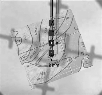
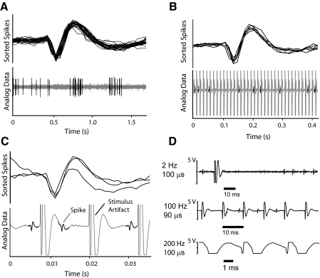
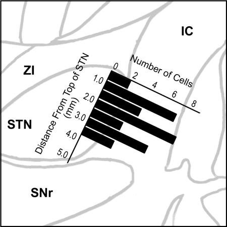
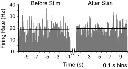
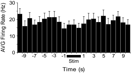
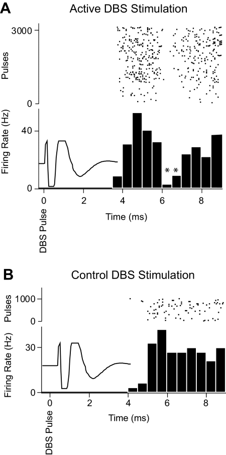
Two broad hypotheses have been advanced to explain the clinical efficacy of deep brain stimulation (DBS) in the subthalamic nucleus (STN) for treatment of Parkinson's disease. One is that stimulation inactivates STN neurons, producing a functional lesion. The other is that electrical stimulation activates the STN output, thus "jamming" pathological activity in basal ganglia-corticothalamic circuits. Evidence consistent with both concepts has been adduced from modeling and animal studies, as well as from recordings in patients. However, the stimulation parameters used in many recording studies have not been well matched to those used clinically. In this study, we recorded STN activity in patients with Parkinson's disease during stimulation delivered through a clinical DBS electrode using standard therapeutic stimulus parameters. A microelectrode was used to record the firing of a single STN neuron during DBS (3-5 V, 80-200 Hz, 90- to 200-micros pulses; 33 neurons/11 patients). Firing rate was unchanged during the stimulus trains, and the recorded neurons did not show prolonged (s) changes in firing rate on termination of the stimulation. However, a brief (approximately 1 ms), short-latency (6 ms) postpulse inhibition was seen in 10 of 14 neurons analyzed. A subset of neurons displayed altered firing patterns, with a predominant shift toward random firing. These data do not support the idea that DBS inactivates the STN and are instead more consistent with the hypothesis that this stimulation provides a null signal to basal ganglia-corticothalamic circuitry that has been altered as part of Parkinson's disease.


Figures

Fig. 1.
Fig. 1.
The physical relationship between the microelectrode and deep brain stimulation (DBS) electrode, enhanced from an intraoperative X-ray and overdrawn on the estimated outline of the subthalamic nucleus (STN). The contact surface of the DBS electrode was positioned ∼1.3 mm posterior to the neuron being recorded with the microelectrode.
Fig. 2.
Fig. 2.
Detectability of action potential waveforms in STN. A: example of spike-sorting based on waveform characteristics. B: example of spikes extracted from the analog signal during a DBS train. C: expanded time base shows spikes between stimulus pulses in the train (same neuron as in B). D: the stimulus artifact changed shape and duration as a function of the stimulus frequency and pulse width. DBS with pulse widths of 100 μs and ∼100 Hz allowed for visible spikes to be detected and sorted during about one half of the time between DBS pulses. Note the complete saturation of the amplifier for several milliseconds during and after each DBS pulse.
Fig. 3.
Fig. 3.
The depth of all cells studied based on microdrive measurements was referenced to the dorsal surface of the STN determined from the microelectrode recordings. The recorded neurons were distributed throughout the dorso-ventral extent of the nucleus. Because of the physical arrangement of the microelectrode anterior to the DBS electrode, the cells were recorded from the anterior portion of the nucleus. IC, internal capsule; SNr, substantia nigra pars reticularis; STN, subthalamic nucleus; ZI, zona incerta.
Fig. 4.
Fig. 4.
Mean firing rate before and after DBS, averaged across all neurons studied for all trials. Firing rate over the 10-s period after stimulus offset was not significantly different from that before stimulus delivery.
Fig. 5.
Fig. 5.
Firing rate during DBS (Stim, bar below trace) was not significantly different from that in the 10 s preceding or after stimulation. Firing during the central 3 s of each stimulus train was used for analysis. Also, because the stimulus artifact precluded detection of spikes for a period during each stimulus duty cycle, firing rate during stimulation was determined using the proportion of time that spikes were detectable. Mean ± SD, 14 neurons, 1-s bins.
Fig. 6.
Fig. 6.
Example of postpulse inhibition of an STN neuron. A: stimulation delivered using contacts 1 and 0 (in STN) resulted in brief (1–2 ms), short-latency (6 ms) inhibition of activity after each pulse in the train. B: control stimulation using the most proximal contacts (3 cathode, 2 anode) did not evoke a postpulse inhibition in this neuron. Stimulus: 100-μs pulses at 100 Hz, 3–5 V. For both A and B, spike times in a 10-ms period aligned with the beginning of the stimulus artifact (superimposed to show time during which spikes were not detectable) were used to construct a pulse-aligned histogram. The spike count in each bin was normalized by the total number of pulses and bin size to give firing rate. Raster record above each histogram shows spike times associated with individual pulses.

References

    1. Albin RL, Young A, Penny JB. The functional anatomy of basal ganglia disorders. Trends Neurosci 12: 366–375, 1989 - PubMed
    1. Benabid AL, Chabardes S, Mitrofanis J, Pollak P. Deep brain stimulation of the subthalamic nucleus for the treatment of Parkinson's disease. Lancet Neurol 8: 67–81, 2009 - PubMed
    1. Bronte-Stewart H, Barberini C, Koop MM, Hill BC, Henderson JM, Wingeier B. The STN beta-band profile in Parkinson's disease is stationary and shows prolonged attenuation after deep brain stimulation. Exp Neurol 215: 20–28, 2009 - PubMed
    1. Butson CR, McIntyre CC. Role of electrode design on the volume of tissue activated during deep brain stimulation. J Neural Eng 3: 1–8, 2006 - PMC - PubMed
    1. Deep Brain Stimulation for Parkinson's Disease Study Group Deep-brain stimulation of the subthalamic nucleus or the pars interna of the globus pallidus in Parkinson's disease. N Engl J Med 345: 956–963, 2001 - PubMed