. 2009 Dec 2;29(48):15266-76.
doi: 10.1523/JNEUROSCI.4650-09.2009.

Rho-associated kinase II (ROCKII) limits axonal growth after trauma within the adult mouse spinal cord

Affiliations

Rho-associated kinase II (ROCKII) limits axonal growth after trauma within the adult mouse spinal cord

Philip Duffy et al. J Neurosci. .

Abstract

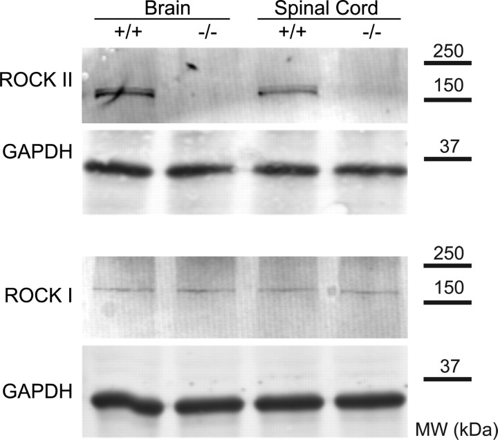
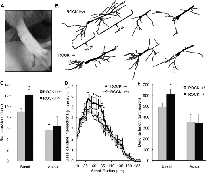
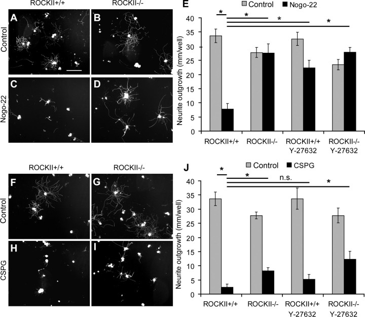
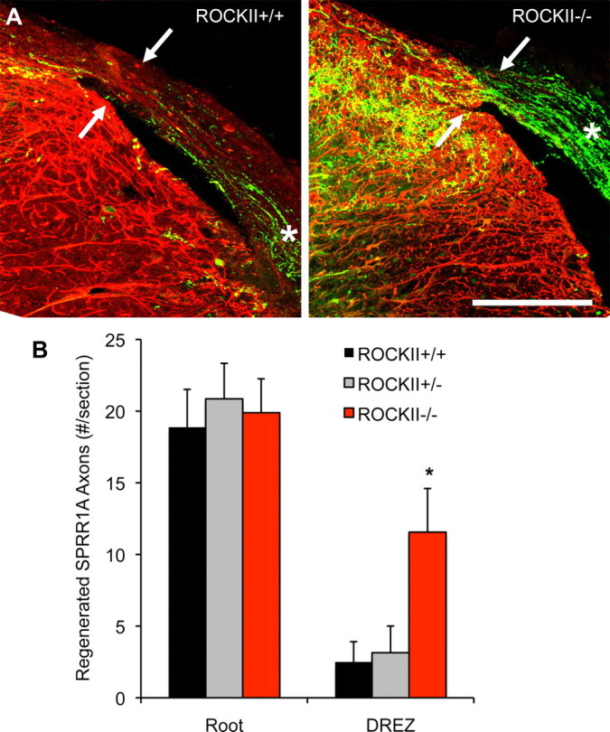
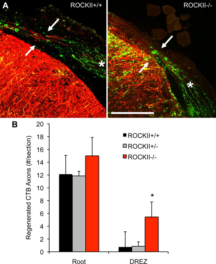
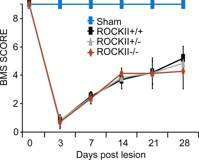
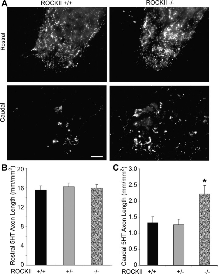
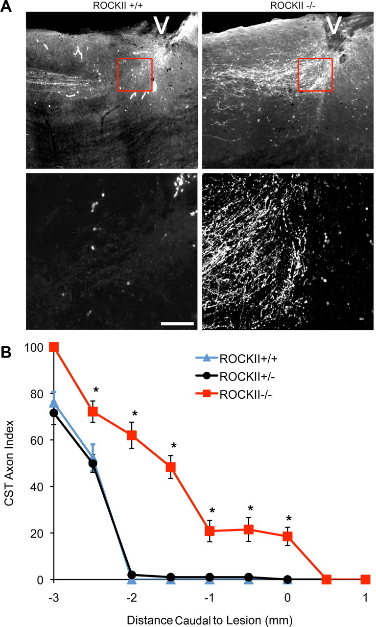
Rho GTPases are thought to mediate the action of several axonal growth inhibitors in the adult brain and spinal cord. RhoA has been targeted pharmacologically in both humans and animals to promote neurite outgrowth and functional recovery following CNS trauma. However, rat spinal cord injury studies suggest a complicated and partial benefit of inhibiting Rho or its downstream effector, Rho-associated kinase (ROCKII). This limited benefit may reflect inhibition of other kinases, poor access, or a minimal role of ROCKII in vivo. Therefore, we studied ROCKII mutant mice to probe this pathway genetically. ROCKII(-/-) dorsal root ganglion neurons are less sensitive to inhibition by Nogo protein or by chondroitin sulfate proteoglycan in vitro. We examined adult ROCKII(-/-) mice in two injury paradigms, cervical multilevel dorsal rhizotomy and midthoracic dorsal spinal cord hemisection. After dorsal root crush injury, the ROCKII(-/-) mice recovered use of the affected forepaw more quickly than did controls. Moreover, multiple classes of sensory axons regenerated across the dorsal root entry zone into the spinal cord of mice lacking ROCKII. After the spinal cord injury, ROCKII(-/-) mice showed enhanced local growth of raphespinal axons in the caudal spinal cord and corticospinal axons into the lesion site. Improved functional recovery was not observed by Basso Mouse Scale score following dorsal hemisection, likely due to developmental defects in the nervous system. Together, these findings demonstrate that the ROCKII gene product limits axonal growth after CNS trauma.


Figures

Figure 1.
Figure 1.
Levels of ROCK protein in ROCKII−/− mice. Brain and spinal cord tissue from ROCKII−/− and wild-type mice was examined by immunoblot for ROCKII and ROCKI protein. A representative blot for each antigen is shown from one pair of three animals with similar results. Anti-GAPDH immunoblots for the same samples demonstrate equal protein loading. Migration of molecular weight (MW) standards is shown at right.
Figure 2.
Figure 2.
ROCKII−/− developmental abnormalities. A, ROCKII−/− pups are frequently born with forepaw and hindpaw digit malformations. B, The number of basal and apical dendrite branchpoints in ROCKII+/+ and ROCKII−/− adult CA1 hippocampal neurons were compared by Golgi staining. C, ROCKII−/− adult mice exhibit a significantly greater number of branchpoints in their basal dendrites compared with ROCKII+/+ littermates. D, E, ROCKII−/− mice also display an increased number of basal dendrite intersections and dendrite length. All data are mean ± SEM, one-way ANOVA, p < 0.05.
Figure 3.
Figure 3.
Nogo-22 and CSPG inhibitory substrates inhibit ROCKII−/− dorsal root ganglion neuronal outgrowth to a lesser extent than ROCKII+/+ neurons. A–J, ROCKII+/+ and ROCKII−/− DRGs were cultured without inhibitor (A, B, F, G), with Nogo-22 (C, D), and with CSPG (H, I) and visualized with anti-βIII tubulin staining. ROCKII+/+ DRG neurite outgrowth was significantly reduced compared with ROCKII−/− neurite outgrowth in the presence of Nogo-22 (E, asterisk). ROCKII+/+ and ROCKII−/− DRG neurite outgrowth was significantly reduced in the presence of CSPG (J, asterisk). However, the effects of CSPG signaling on neuronal outgrowth were less inhibitory for ROCKII−/− neurons than for ROCKII+/+ DRGs (J, asterisk). Addition of soluble Y-27632 negated the inhibitory effects of Nogo-22 on ROCKII+/+ DRG neurite outgrowth and, to a lesser extent, CSPGs on ROCKII+/+ and ROCKII−/− DRG neurite outgrowth (E, J). Scale bar: (in A) A–I, 50 μm. All data are mean ± SEM, one-way ANOVA, p < 0.05.
Figure 4.
Figure 4.
ROCKII gene deletion promotes regeneration of SPRR1A-expressing sensory neurons after rhizotomy. A, Photomicrographs illustrate high-power transverse sections of cervical spinal cord from adult ROCKII+/+ (left) and ROCKII−/− (right) mice which underwent dorsal rhizotomy at cervical spinal cord segments C5–C8 28 d previously. SPRR1A-immunoreactive axons (green channel) can be seen in the dorsal root of both genotypes (asterisk). However, significantly greater numbers of axons are present entering the DREZ in the ROCKII−/− group (arrow in right photomicrograph) than in control group (arrow in left photomicrograph, quantified in B, asterisk). Reactive CNS tissue is visualized with anti-GFAP immunofluorescence (red channel). Scale bar, 100 μm. Mean ± SEM, one-way ANOVA, p < 0.05.
Figure 5.
Figure 5.
ROCKII gene deletion promotes regeneration of CGRP-expressing sensory neurons after rhizotomy. A, Photomicrographs illustrate high-power transverse sections of cervical spinal cord from adult ROCKII+/+ (left) and ROCKII−/− (right) mice which underwent dorsal rhizotomy at cervical spinal cord segments C5–C8 28 d previously. CGRP-immunoreactive axons can be seen in the dorsal root of both genotypes (asterisk, both photomicrographs). However, significantly greater numbers of axons are present entering the DREZ in the ROCKII−/− group (arrow in right photomicrograph) than in control group (arrow in left photomicrograph, quantified in B, asterisk). Anti-GFAP immunofluorescence is shown in the red channel. Scale bar, 100 μm. Mean ± SEM, one-way ANOVA, p < 0.05.
Figure 6.
Figure 6.
ROCKII gene deletion promotes regeneration of CTB-labeled sensory neurons after rhizotomy. A, Photomicrographs illustrate high-power transverse sections of cervical spinal cord from adult ROCKII+/+ (left) and ROCKII−/− (right) mice which underwent dorsal rhizotomy at cervical spinal cord segments C5–C8 28 d previously. CTB-labeled axons (green channel) can be seen in the dorsal root of both genotypes (asterisk, both photomicrographs). However, significantly greater numbers of axons are present entering the DREZ in the ROCKII−/− group (arrow in right photomicrograph) than in control group (arrow in left photomicrograph, quantified in B, asterisk). Reactive astrocytes are visualized by anti-GFAP immunofluorescence (red channel). Scale bar, 100 μm. Mean ± SEM, one-way ANOVA, p < 0.05.
Figure 7.
Figure 7.
ROCKII−/− behavioral recovery following cervical rhizotomy. A, Prelesion, forelimb use during rearing is not significantly different between ROCKII+/+, ROCKII+/−, and ROCKII−/− groups. B, Following injury, ROCKII−/− mice display a reduced dependency on uninjured paw for exploration during a rear by 3 d postlesion (asterisk). This trend is continued until 15 d postlesion, by which time groups are not significantly different from each other. C, ROCKII−/− mice do not display enhanced recovery of thermal sensation following rhizotomy. Withdrawal times after the thermal stimulus were not significantly difference between animal groups at any time point. Injured groups showed decreased withdrawal latencies at early time points postinjury. This trend was not present at >5 d postinjury, when animals in each injured group had become hypersensitive to the thermal stimulus. Hypersensitivity continued throughout the remainder of the testing period. Mean ± SEM, 1 WAY ANOVA, p < 0.05.
Figure 8.
Figure 8.
ROCKII−/− mice do not display enhanced recovery in the Basso Mouse Scale open field locomotor test following hemisection. ROCKII+/+ (black), ROCKII+/− (brown), ROCKII−/− (red), and sham (gray, genotypes combined) mice underwent open field locomotor assessment after hemisection lesion. ROCKII+/+ (black), ROCKII+/− (brown), and ROCKII−/− (red) groups all exhibited significant deficits in BMS scores following injury compared with sham (gray) animals, which was maintained throughout the testing period (asterisk). No statistical difference was observed between injured groups at any time point. Mean ± SEM.
Figure 9.
Figure 9.
ROCKII−/− raphespinal fiber length is increased caudal to a hemisection. A, Photomicrographs of 5-HT-immunopositive serotonergic raphespinal tract neurons in transverse sections of ventral horn gray matter rostral (top panel) and caudal (second panel) to a hemisection lesion. B, Raphespinal tract fiber lengths are not significantly different between ROCKII+/+, ROCKII+/−, and ROCKII−/− animals rostral to a lesion. C, Caudal to a lesion, ROCKII−/− animals exhibit significantly greater fiber length compared with ROCKII+/+ and ROCKII+/− mice (asterisk). Mean ± SEM, one-way ANOVA, p < 0.05. Scale bar 100 μm.
Figure 10.
Figure 10.
ROCKII gene deletion increases the number of CST axons in the astrocytic scar of an SCI. A, Photomicrographs illustrate sagittal sections of ROCKII+/+ (left) and ROCKII−/− (right) mice 6 weeks after hemisection. BDA-immunoreactive CST axons can be seen approaching the lesion site (indicated by the white arrowheads) in both genotypes. High-power magnifications of the lesion area (highlighted boxes in top, shown in bottom) show regenerating CST axons at distances >3 mm rostral from the lesion epicenter in ROCKII+/+ mice (lower left photomicrograph), whereas ROCKII−/− CST axons are present entering the lesion site (lower right photomicrograph). Scale bar, Top, 800 μm; bottom, 200 μm. B, Quantification of CST axon growth illustrates that significantly greater numbers of axons are present at various points leading up to the lesion epicenter (mean ± SEM, 1-way ANOVA, p < 0.05) in ROCKII−/− animals (red) compared with ROCKII+/− (black) and ROCKII+/+ (gray) mice.

References

    1. Abad F, Feria M, Boada J. Chronic amitriptyline decreases autotomy following dorsal rhizotomy in rats. Neurosci Lett. 1989;99:187–190. - PubMed
    1. Basso DM, Fisher LC, Anderson AJ, Jakeman LB, McTigue DM, Popovich PG. Basso Mouse Scale for locomotion detects differences in recovery after spinal cord injury in five common mouse strains. J Neurotrauma. 2006;23:635–659. - PubMed
    1. Benson MD, Romero MI, Lush ME, Lu QR, Henkemeyer M, Parada LF. Ephrin-B3 is a myelin-based inhibitor of neurite outgrowth. Proc Natl Acad Sci U S A. 2005;102:10694–10699. - PMC - PubMed
    1. Bonilla IE, Tanabe K, Strittmatter SM. Small proline-rich repeat protein 1A is expressed by axotomized neurons and promotes axonal outgrowth. J Neurosci. 2002;22:1303–1315. - PMC - PubMed
    1. Borisoff JF, Chan CC, Hiebert GW, Oschipok L, Robertson GS, Zamboni R, Steeves JD, Tetzlaff W. Suppression of Rho-kinase activity promotes axonal growth on inhibitory CNS substrates. Mol Cell Neurosci. 2003;22:405–416. - PubMed

Publication types

MeSH terms