. 2009 Dec 2;29(48):15323-30.
doi: 10.1523/JNEUROSCI.2107-09.2009.

Estradiol-induced estrogen receptor-alpha trafficking

Affiliations

Estradiol-induced estrogen receptor-alpha trafficking

Galyna Bondar et al. J Neurosci. .

Abstract

Estradiol has rapid actions in the CNS that are mediated by membrane estrogen receptors (ERs) and activate cell signaling pathways through interaction with metabotropic glutamate receptors (mGluRs). Membrane-initiated estradiol signaling increases the free cytoplasmic calcium concentration ([Ca(2+)](i)) that stimulates the synthesis of neuroprogesterone in astrocytes. We used surface biotinylation to demonstrate that ERalpha has an extracellular portion. In addition to the full-length ERalpha [apparent molecular weight (MW), 66 kDa], surface biotinylation labeled an ERalpha-immunoreactive protein (MW, approximately 52 kDa) identified by both COOH- and NH(2)-directed antibodies. Estradiol treatment regulated membrane levels of both proteins in parallel: within 5 min, estradiol significantly increased membrane levels of the 66 and 52 kDa ERalpha. Internalization, a measure of membrane receptor activation, was also increased by estradiol with a similar time course. Continuous treatment with estradiol for 24-48 h reduced ERalpha levels, suggesting receptor downregulation. Estradiol also increased mGluR1a trafficking and internalization, consistent with the proposed ERalpha-mGluR1a interaction. Blocking ER with ICI 182,780 or mGluR1a with LY 367385 prevented ERalpha trafficking to and from the membrane. Estradiol-induced [Ca(2+)](i) flux was also significantly increased at the time of peak ERalpha activation/internalization. These results demonstrate that ERalpha is present in the membrane and has an extracellular portion. Furthermore, membrane levels and internalization of ERalpha are regulated by estradiol and mGluR1a ligands. The pattern of trafficking into and out of the membrane suggests that the changing concentration of estradiol during the estrous cycle regulates ERalpha to augment and then terminate membrane-initiated signaling.


Figures

Figure 1.
Figure 1.
Postpubertal hypothalamic astrocytes were incubated with vehicle (0 min) or in the presence of 1 nm estradiol for 5 and 30 min and 1, 2, 24, and 48 h. Astrocytes were then biotinylated, the biotin was removed, and the labeled proteins were separated and detected with ERα, GPR30, and LIM K1 antibodies. A, Two ERα-immunoreactive (ERα-i) bands were identified: 66 and 52 kDa. The cytoplasmic protein LIM K1 and the putative membrane estrogen receptor GPR30 were not labeled with surface biotinylation. B, Estradiol treatment increased both the 66 and 52 kDa ERα-i bands. In the first lane, cells were not surface biotinylated (no biotin), thus no biotinylated ERα-i were labeled. Detection of the 66 kDA ERα required a 2 h exposure compared with a 1–2 min exposure for the 52 kDa ERα-i. C, Quantification of the 66 and 52 kDa ERα-i bands was calculated by comparing optical density of the ERα-i bands with the optical density of the β-actin bands. Both 66 and 52 kDa ERα-i bands are regulated in parallel by estradiol treatment, but the amount of 66 kDa ERα was much less at each time point. Data are mean ± SEM (n = 4). *,+Statistical differences at the p < 0.05 level compared with 0 min for each molecular-weight species. E2, Estradiol.
Figure 2.
Figure 2.
A, Estradiol treatment transiently increases membrane ERα-immunoreactive (ERα-i) bands in postpubertal hypothalamic astrocytes. Basal levels of ERα-i bands were observed before estradiol treatment (0 min). These levels were rapidly increased (5 min time point) (*p < 0.05), with a maximum at 30 min (*p < 0.05) and a slight depression after 1 h (*p < 0.05), and remained elevated for 2 h of estradiol stimulation (*p < 0.05). After 24–48 h of estradiol treatment, ERα-i bands returned to basal levels (p > 0.05). GPR30 was not biotinylated on the surface despite incubating the astrocytes with estradiol for up to 48 h. B, To track internalization, astrocytes were biotinylated and treated with estradiol, and the biotin was stripped from the surface of the cells with glutathione. Under these conditions, ERα-i bands are biotinylated, but after glutathione treatment, the only biotinylated receptors remaining are those that were internalized. The time course of internalization matched the time course of the estradiol-induced trafficking to the membrane. In the first lane, the biotin was not removed by glutathione (nonstripped). The amount of internalized ERα-i bands, with varying estradiol treatment, began increasing at 5 min (*p < 0.05) and reached its maximum at 30 min to 1 h (*p < 0.05). After 2 h of estradiol incubation, the level of internalized ERα decreased compared with the maximum but was still statistically significant from the 0 min time point (*p < 0.05). At the 24–48 h time points, internalized ERα-i levels reached basal levels comparable to 0 min (p > 0.05). All the data are mean ± SEM (n = 4). *Statistical differences at the p < 0.05 level compared with 0 min for each experiment. E2, Estradiol.
Figure 3.
Figure 3.
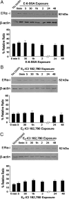
A, Postpubertal hypothalamic astrocytes were incubated with 1 nm E-6-BSA before biotinylation. The membrane-constrained estradiol did not change membrane levels of ERα-immunoreactive (ERα-i) bands, indicating that estradiol acts within the cell to regulate ERα-i band trafficking to the membrane. B, To study the effect of estradiol on ERα-i band trafficking to the membrane, cells were preincubated with charcoal-stripped medium containing 1 μm ICI 182,780, an ER antagonist, for 1 h before and during 1 nm estradiol treatment. ICI 182,780 prevented trafficking of ERα-i bands to the membrane. C, To study the effect of estradiol on ERα-i band internalization, cells were surface biotinylated and treated with estradiol (1 nm) and ICI 182,780 (1 μm), which prevented internalization of ERα-i band. All the data are mean ± SEM (n = 4). *Differences at the p < 0.05 level compared with 0 min for each experiment. E2, Estradiol.
Figure 4.
Figure 4.
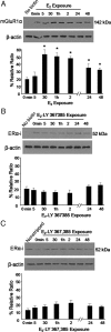
A, To detect whether estradiol regulates mGluR1a trafficking, postpubertal hypothalamic astrocytes were treated with estradiol (1 nm) for the indicated times and surface biotinylated. Levels of membrane mGluR1a were increased by estradiol treatment. By 30 min, mGluR1a levels were significantly higher than no previous estradiol treatment (0 min; *p < 0.05). Membrane levels of mGluR1a did not return to basal levels during the experiment (48 h). B, The mGluR1a antagonist LY 367385 (50 μm) blocked the estradiol-induced ERα-immunoreactive (ERα-i) trafficking to the membrane. C, Similarly, 50 μm LY 367385 prevented the estradiol-induced ERα-i internalization. All the data are mean ± SEM (n = 4). *Differences at the p < 0.05 level compared with 0 min for each experiment. E2, Estradiol.
Figure 5.
Figure 5.
A, Duration of steroid-starvation increased the estradiol-induced [Ca2+]i flux in postpubertal hypothalamic astrocytes. Estradiol at 10 nm induced a greater [Ca2+]i response after a 6 h (*p < 0.001) and 12 h (*p < 0.001) steroid-starvation compared with no prior (0 h) steroid-starvation. B, Prior estradiol exposure transiently increased [Ca2+]i flux in astrocytes. After a 12 h steroid-starvation without previous estradiol incubation (0 min), estradiol at 10 nm induced a robust [Ca2+]i flux. The 10 nm estradiol-induced [Ca2+]i flux increased after exposure to 1 nm estradiol for 30 min (*p < 0.05). However, incubation with 1 nm estradiol for 5 min, 1 h, or 2 h (p > 0.05) was no different than no prior incubation with estradiol (0 min). All the data are mean ± SEM. *Differences at the p < 0.05 level compared with 0 min.

Similar articles

Cited by

References

    1. Acconcia F, Ascenzi P, Bocedi A, Spisni E, Tomasi V, Trentalance A, Visca P, Marino M. Palmitoylation-dependent estrogen receptor alpha membrane localization: regulation by 17beta-estradiol. Mol Biol Cell. 2005;16:231–237. - PMC - PubMed
    1. Azcoitia I, Sierra A, Garcia-Segura LM. Localization of estrogen receptor beta-immunoreactivity in astrocytes of the adult rat brain. Glia. 1999;26:260–267. - PubMed
    1. Beyer C, Pawlak J, Karolczak M. Membrane receptors for oestrogen in the brain. J Neurochem. 2003;87:545–550. - PubMed
    1. Bollig A, Miksicek RJ. An estrogen receptor-alpha splicing variant mediates both positive and negative effects on gene transcription. Mol Endocrinol. 2000;14:634–649. - PubMed
    1. Boulware MI, Weick JP, Becklund BR, Kuo SP, Groth RD, Mermelstein PG. Estradiol activates group I and II metabotropic glutamate receptor signaling, leading to opposing influences on cAMP response element-binding protein. J Neurosci. 2005;25:5066–5078. - PMC - PubMed

Publication types

MeSH terms

LinkOut - more resources