. 2009 Dec 15;106(50):21425-30.
doi: 10.1073/pnas.0912021106. Epub 2009 Dec 2.

Activity of guard cell anion channel SLAC1 is controlled by drought-stress signaling kinase-phosphatase pair

Affiliations

Activity of guard cell anion channel SLAC1 is controlled by drought-stress signaling kinase-phosphatase pair

Dietmar Geiger et al. Proc Natl Acad Sci U S A. .

Abstract

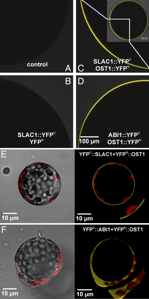
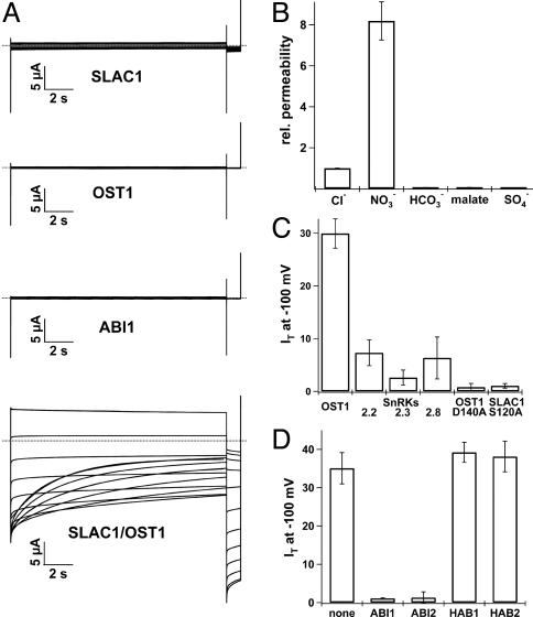
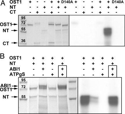
In response to drought stress the phytohormone ABA (abscisic acid) induces stomatal closure and, therein, activates guard cell anion channels in a calcium-dependent as well as-independent manner. Two key components of the ABA signaling pathway are the protein kinase OST1 (open stomata 1) and the protein phosphatase ABI1 (ABA insensitive 1). The recently identified guard cell anion channel SLAC1 appeared to be the key ion channel in this signaling pathway but remained electrically silent when expressed heterologously. Using split YFP assays, we identified OST1 as an interaction partner of SLAC1 and ABI1. Upon coexpression of SLAC1 with OST1 in Xenopus oocytes, SLAC1-related anion currents appeared similar to those observed in guard cells. Integration of ABI1 into the SLAC1/OST1 complex, however, prevented SLAC1 activation. Our studies demonstrate that SLAC1 represents the slow, deactivating, weak voltage-dependent anion channel of guard cells controlled by phosphorylation/dephosphorylation.


Conflict of interest statement

The authors declare no conflict of interest.

Figures

Fig. 1.
Fig. 1.
ABA activation of slow anion channels in A. thaliana guard cell protoplasts. (A) Representative macroscopic current responses from WT, ost1–2, and slac1–3 to voltage pulses in the range from + 44 mV to −136 mV are shown. The zero-current level is indicated by the dotted line. (B) Steady-state current densities plotted against the clamped voltages. Data points represent the mean ± SE. The number of protoplasts studied in at least 3 independent experiments were in (B) n = 11 for ost1–2, n = 5 for slac1–3, and n = 12 for WT. The experiments from (A) and (B) were performed in the presence of 25 μM ABA and 110 nM cytosolic free Ca2+.
Fig. 2.
Fig. 2.
Interactions between proteins involved in the ABA signaling pathway by bimolecular fluorescence complementation (BIFC) in Xenopus oocytes (A–D) as well as Arabidopsis mesophyll protoplasts (E) and (F). Pictures (A–D), taken with a confocal laser scanning microscope, show a quarter of an optical slice of an oocyte (see inset in (C). (A) Noninjected control oocyte. (B) SLAC1::YFPC coexpressed with YFPN. (C) SLAC1::YFPC coexpressed with OST1::YFPN. (D) ABI1::YFPC coexpressed with OST1::YFPN. (E and F) Interaction of SLAC1 and OST1, as well as ABI1 and OST1, monitored in transiently transformed Arabidopsis mesophyll protoplasts by bimolecular fluorescence complementation. (Left) Overlay of transmitted light and chlorophyll fluorescence. (Right) Overlay of YFP and chlorophyll fluorescence. Plasmid combinations were as follows: (E) YFPC::SLAC1 plus YFPN::OST1 and (F) YFPC::ABI1 plus YFPN::OST1.
Fig. 3.
Fig. 3.
Regulation of SLAC1 activity by distinct kinases and phosphatases. (A) Whole-oocyte current recordings in standard bath solution (30 mM Cl, pH 5.6) upon 15 s voltage pulses ranging from + 40 to −180 mV in 20 mV decrements followed by a 3 s voltage pulse to −120 mV. The holding potential was 0 mV. No prominent current responses of oocytes expressing SLAC1, OST1, or ABI1 alone were recorded (Upper). Only coexpression of SLAC1 together with OST1 (Lower) resulted in macroscopic anion currents slowly deactivating at negative membrane potentials. Representative cells are shown. (B) Relative permeability of SLAC1 for physiological relevant anions (permeability for Cl was set to 1). Standard bath solution contained 50 mM of the respective anion, pH 5.6 (n = 5). (C) Coexpression studies in oocytes elucidated the specificity of SnRKs toward SLAC1. Instantaneous SLAC1 currents (IT in μA) activated at −100 mV in standard bath solution are shown. Among the tested SnRKs, OST1 coexpression with SLAC1 resulted in the largest anion currents. The coexpression of SLAC1 together with the inactive OST1 mutant D140A prevented SLAC1 currents completely (n ≥ 5). (D) ABI1 and ABI2 coexpression inhibited SLAC1 activation by OST1. Coinjection of HAB1 and HAB2, however, could not prevent SLAC1/OST1 mediated anion currents (n ≥ 3 in μA). Experiments with SLAC1 activated by SnRK/OST1 were performed with oocytes expressing SLAC1::YFPC and SnRK/OST1::YFPN. Error bars in (B–C) represent SD.
Fig. 4.
Fig. 4.
In vitro kinase activity of native recombinant GST1-tagged proteins. (A) Phosphorylation of SLAC1 termini (NT and CT) by OST1 was tested by using radio-labeled [γ32] ATP (Left: Coomassie stained SDS PAGE; Right: radioautogram of the gel; the presence of proteins in the reaction assay was indicated by +). Only SLAC1 NT was phosphorylated by OST1. In contrast to OST1 WT, the OST1 mutant D140A did not phosphorylate SLAC1 NT. Arrowheads indicate the position of recombinant proteins. (B) In vitro kinase assays with native recombinant proteins revealed that OST1 activity was prevented by ABI1 and the ATP analogue ATPγS. When SLAC1 NT was phosphorylated before adding ABI1 together with ATPγS (indicated by the black box, lane 4) we could not detect any dephosphorylation activity of ABI1 within 45 min of incubation at RT. The molecular weight of the protein ladder is indicated in kDa.

Similar articles

Cited by

References

    1. Assmann SM, Shimazaki K. The multisensory guard cell. Stomatal responses to blue light and abscisic acid. Plant Physiol. 1999;119:809–816. - PMC - PubMed
    1. Hetherington AM, Woodward FI. The role of stomata in sensing and driving environmental change. Nature. 2003;424:901–908. - PubMed
    1. Raschke K. Action of abscisic acid on guard cells. In: Zeiger GDF E, Cowan IR, editors. Stomatal Funktion. Stanford, CA: Stanford Univ Press; 1987. pp. 253–279.
    1. Roelfsema MR, Hedrich R. In the light of stomatal opening: New insights into “the Watergate”. New Phytol. 2005;167:665–691. - PubMed
    1. Schroeder JI, Kwak JM, Allen GJ. Guard cell abscisic acid signalling and engineering drought hardiness in plants. Nature. 2001;410:327–330. - PubMed

Publication types