. 2009 Dec 22;106(51):21789-94.
doi: 10.1073/pnas.0912152106. Epub 2009 Dec 2.

IL-17-producing T cells can augment autoantibody-induced arthritis

Affiliations

IL-17-producing T cells can augment autoantibody-induced arthritis

Jonathan P Jacobs et al. Proc Natl Acad Sci U S A. .

Abstract

Rheumatoid arthritis is a T lymphocyte-mediated disorder, but the precise nature of T cell involvement remains unclear. In the K/BxN mouse model of inflammatory arthritis, T cells initiate disease by providing help to B cells to produce arthritogenic autoantibodies. Here, we have characterized an additional, nonhumoral role for T cells in promoting autoantibody-induced arthritis. Autoreactive KRN T cells introduced either by direct transfer or bone marrow transplantation into B-cell-deficient hosts enhanced K/BxN serum-transferred arthritis, an effect that was dependent on expression of the cognate MHC-molecule/peptide complex. The T cell influence was dependent on interleukin (IL)-17; in contrast, standard serum-transferred arthritis, unenhanced by the addition of T cells, was unaffected by IL-17 neutralization. An IL-17-producing population of transferred KRN T cells was identified and found to be supported by the cotransfer of arthritogenic autoantibodies. IL-17-producing KRN T cells were enriched in inflamed joints of K/BxN mice, suggesting either selective recruitment or preferential differentiation. These results demonstrate the potential for autoreactive T cells to play two roles in the development of arthritis, both driving the production of pathogenic autoantibodies and bolstering the subsequent inflammatory cascade dependent on the innate immune system.


Conflict of interest statement

The authors declare no conflict of interest.

Figures
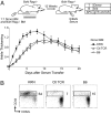
Fig. 1.
Fig. 1.
KRN T cells augment serum-transferred arthritis. Irradiated BxN Rag−/− mice were reconstituted with a 1:1 mix of KRN (n = 6), OTII (Ctl TCR, n = 6), or B6 (n = 3) BM and BxN Rag−/− BM. (A) Serum-transferred arthritis was induced after 12 weeks and assessed by ankle thickening (in millimeters), represented as mean change from day 0 ± SEM. P = 0.002 for KRN vs. OTII using area under the curve analysis. A side table summarizes the source of T cells (T) and the MHC genotypes (b/b for B6, g7/b for BxN) of B cells (B) and APCs in recipient mice. (B) Donor-derived CD4+ T cells from cervical lymph nodes surgically removed at week 11 were stained for CD44 and CD62L.
Fig. 2.
Fig. 2.
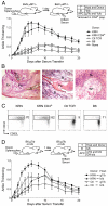
CD4+ KRN T cells short-term activated by their cognate MHC-molecule/peptide complex boost serum-transferred arthritis. (A–C) Twenty million splenocytes from KRN, OTII (Ctl TCR), or B6 mice were transferred into BxN μMT−/− recipients (n = 5). An additional group received KRN CD4+ T cells prepared from 2 × 10⁷ KRN splenocytes enriched by magnetic separation to 80–85% purity. One control group received medium alone (None). (A) Arthritis was induced in recipient mice 1 day after cell transfer and measured by ankle thickening. P = 3 × 10⁻⁵ for KRN vs. Ctl TCR, P = 0.008 for KRN CD4+ vs. Ctl TCR, and P = 0.43 for KRN vs. KRN CD4+. (B) Additional BxN μMT−/− mice receiving KRN splenocytes and K/BxN serum were killed on day 5 of arthritis. H&E staining demonstrated extensive leukocyte infiltration, synovial hyperplasia with pannus (Pa) formation, and fibrinous exudates (Ex) (Left magnification, ×40); pannus invasion into cartilage (C) and superficial bone (B) (Middle magnification, ×200); and neutrophil accumulation in synovial lining (SL) and synovial fluid (SF) (Right magnification, ×400). (C) Transferred CD4+ T cells (identified by the absence of NOD-derived CD45.1) were isolated from cervical lymph nodes on day 5 and stained for CD44 and CD62L. (D) Twenty million splenocytes from KRN or OTII (Ctl) mice were transferred into B6.H2g7/b μMT−/− or B6.H2b/b μMT−/− littermates (n = 6). Arthritis was induced 1 day after cell transfer with a limiting dose of K/BxN serum. P = 0.02 for KRN→g7/b vs. KRN→b/b.
Fig. 3.
Fig. 3.
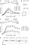
Monoclonal T cell autoreactivity is not sufficient to augment serum-transferred arthritis. (A and B) Twenty million splenocytes from AND/CD45.1 B10.BR (AND+) or B10.BR (AND) mice were transferred into Eα-MCC (MCC+) or transgene-negative B10.BR littermates (MCC) (n = 7 AND+→MCC+, n = 5 for other groups). (A) Arthritis was induced in recipient mice 1 day after cell transfer with a limiting dose of K/BxN serum and measured by ankle thickening. P = 0.008 for AND→MCC+ vs. AND→MCC. (B) Transferred CD4+ T cells (CD45.1+) obtained from cervical lymph nodes surgically removed on day 5 of arthritis were stained for CD44 and CD62L. (C and D) Twenty million splenocytes from KRN or OTII were transferred into Act-mOva/BxN μMT−/− (OVA+) or BxN μMT−/− littermates (OVA) (n = 5). (C) Arthritis was induced in recipient mice 1 day after cell transfer and measured by ankle thickening. (D) Transferred CD4+ T cells (CD45.1) obtained from cervical lymph nodes surgically removed on day 5 of arthritis were stained for CD44 and CD62L.
Fig. 4.
Fig. 4.
Augmentation of serum-transferred arthritis requires IL-17 but not IL-6, IL-12p40, or KRN-T cell-derived IFNγ, TNFα, and IL-4. (A–C) Serum-transferred arthritis was induced in BxN μMT−/− mice receiving splenocytes from (A) IFNγ+/− or IFNγ−/− (n = 5) KRN littermates or OTII mice (Ctl). Similar comparisons were made of (B) TNFα+/− vs. TNFα−/− (n = 5) and (C) IL-4+/− vs. IL-4−/− (n = 9) KRN donors. (D) 100 μg anti-IL-17 (αIL-17) or isotype control (IgG2a) antibodies were given i.p. on days 0, 2, and 4 (indicated by arrows) of serum-transferred arthritis in BxN μMT−/− mice receiving KRN splenocytes (n = 5). OTII recipients were used as a control. P = 0.004 for KRN αIL-17 vs. KRN IgG2a; P = 0.35 for KRN αIL-17 vs. Ctl. (E) B6 mice with serum-transferred arthritis were treated with 100 μg αIL-17 or isotype control IgG2a antibodies on days 0, 2, and 4 (n = 6). (F) As in (D) except that KRN recipients were treated with 500 μg anti-IL-12p40 (αIL-12p40), 2.66 mg/kg anti-IL-6 (αIL-6), or 500 μg Rat IgG control (IgG) antibodies. A representative experiment (n = 4) of three is shown. P = 0.003 for KRN αIL-12p40 vs. KRN IgG.
Fig. 5.
Fig. 5.
Transferred IL-17-producing CD4+ KRN T cells are supported by arthritogenic antibodies. (A and B) K/BxN serum or PBS was injected i.p. into BxN μMT−/− mice 1 and 3 days after transfer of KRN or OTII (Ctl) splenocytes. Six days after cell transfer, cervical and inguinal lymph nodes were harvested for flow cytometry. (A) Intracellular staining for IL-17 and IFNγ is shown for transferred CD4+ T cells. (B) The bar graphs depict the mean percentage ± SEM of transferred CD4+ T cells in each group expressing IL-17 or IFNγ (n = 5 for KRN groups, n = 4 for Ctl groups). (C) Intracellular staining for IL-17 and surface staining for CCR6, a chemokine receptor expressed on Th17 cells, is shown for CD4+ T cells from donor KRN B6 splenocytes and splenocytes obtained from B6 mice on day 5 of serum-transferred arthritis.
Fig. 6.
Fig. 6.
IL-17-producing CD4+ KRN T cells are enriched in K/BxN transgenic mice, with greatest concentration in arthritic joints. (A) Intracellular staining for IL-17 and IFNγ is shown for CD4+ T cells from the spleen (SPL), popliteal lymph nodes (PLN), and ankle synovial fluid (SF) (K/BxN only) of 8-week-old K/BxN mice and transgene negative littermates. (B) The bar graphs depict the mean percentage ± SEM of CD4+ T cells that express IL-17 in each group (n = 4).

Similar articles

Cited by

References

    1. Lundy SK, Sarkar S, Tesmer LA, Fox DA. Cells of the synovium in rheumatoid arthritis. T lymphocytes. Arthritis Res Ther. 2007;9:202. - PMC - PubMed
    1. Skapenko A, Leipe J, Lipsky PE, Schulze-Koops H. The role of the T cell in autoimmune inflammation. Arthritis Res Ther 7 Suppl. 2005;2:S4–14. - PMC - PubMed
    1. Monach PA, Benoist C, Mathis D. The role of antibodies in mouse models of rheumatoid arthritis, and relevance to human disease. Adv Immunol. 2004;82:217–248. - PubMed
    1. Kouskoff V, et al. Organ-specific disease provoked by systemic autoimmunity. Cell. 1996;87:811–822. - PubMed
    1. Sawa S, et al. Autoimmune arthritis associated with mutated interleukin (IL)-6 receptor gp130 is driven by STAT3/IL-7-dependent homeostatic proliferation of CD4+ T cells. J Exp Med. 2006;203:1459–1470. - PMC - PubMed

Publication types