. 2009 Dec 22;106(51):21579-84.
doi: 10.1073/pnas.0910708106. Epub 2009 Dec 2.

The crystal structure of apo-FtsH reveals domain movements necessary for substrate unfolding and translocation

Affiliations

The crystal structure of apo-FtsH reveals domain movements necessary for substrate unfolding and translocation

Christoph Bieniossek et al. Proc Natl Acad Sci U S A. .

Abstract

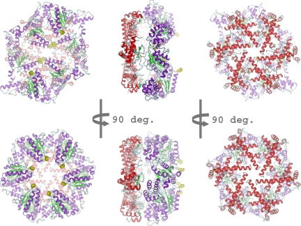
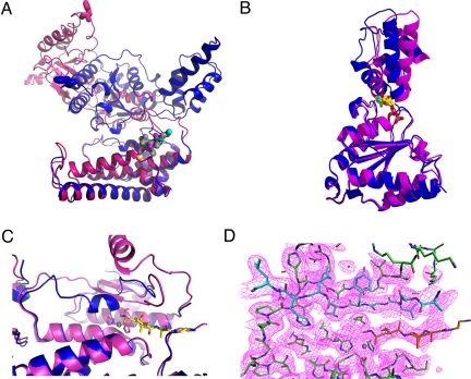
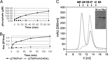
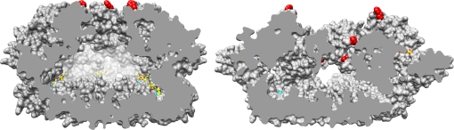
The hexameric membrane-spanning ATP-dependent metalloprotease FtsH is universally conserved in eubacteria, mitochondria, and chloroplasts, where it fulfills key functions in quality control and signaling. As a member of the self-compartmentalizing ATPases associated with various cellular activities (AAA+ proteases), FtsH converts the chemical energy stored in ATP via conformational rearrangements into a mechanical force that is used for substrate unfolding and translocation into the proteolytic chamber. The crystal structure of the ADP state of Thermotoga maritima FtsH showed a hexameric assembly consisting of a 6-fold symmetric protease disk and a 2-fold symmetric AAA ring. The 2.6 A resolution structure of the cytosolic region of apo-FtsH presented here reveals a new arrangement where the ATPase ring shows perfect 6-fold symmetry with the crucial pore residues lining an open circular entrance. Triggered by this conformational change, a substrate-binding edge beta strand appears within the proteolytic domain. Comparison of the apo- and ADP-bound structure visualizes an inward movement of the aromatic pore residues and generates a model of substrate translocation by AAA+ proteases. Furthermore, we demonstrate that mutation of a conserved glycine in the linker region inactivates FtsH.


Conflict of interest statement

The authors declare no conflict of interest.

Figures

Fig. 1.
Fig. 1.
Cartoon representation of ADP-FtsH (Top) and apo-FtsH (Bottom) with view onto AAA ring (Left), side (Middle) and protease (Right). The protease ring is shown in red and the AAA ring in magenta and green. Zinc ions are displayed as cyan spheres, the pore residues Phe-234 in yellow space-filling mode. Carbon atoms of ADP and TAPI are shown in gray. (A) ADP-bound state, the AAA ring has C2 symmetry. (B) Apo-state, the AAA ring has C6 symmetry.
Fig. 2.
Fig. 2.
(A) Overlay of monomers in the ADP (blue) and apo-state (magenta). The pore phenylalanine Phe-234 is shown as sticks. The distance of these residues between ADP and apo-form is about 45 Å. Zn+ is shown as yellow sphere, the hydroxamate inhibitor as stick model with gray carbon atoms. The protease domain (bottom) was used as reference for the alignment. (B) Overlay of the AAA domain only. The angle between “wedge” subdomain and α-helical subdomain increases by about 20° in the apo form (red) compared to the ADP form (blue). (C) Overlay of the proteolytic active site in the ADP (blue) and apo-state (magenta). The active-size switch (residues 450 to 460) adopts a helical conformation in the ADP state and forms a β-strand in the apo-state. TAPI is displayed with yellow carbon atoms, Zn2+ as gray sphere. (D) Active site with electron density (1.0 σ) above mean. Zn2+ is shown as gray sphere, the active-site switch (residue 450 to 460) is depicted in cyan. The TAPI inhibitor is shown with orange carbon atoms.
Fig. 3.
Fig. 3.
Activity assays and oligomeric state for the G404L mutant. (A) ATPase activities of wild-type Δ(tm)FtsH (solid line, ●) and Δ(tm)G404L (dashed line, ▴). (B) Resorufin casein proteolytic assay, symbols as in (A). (C) Gel filtration profile of wild-type Δ(tm)FtsH (solid line) and G404L (dashed line). The insert shows the SDS/PAGE with molecular weight markers in lane M (66, 45, 24, 14, and 12 kDa), wild-type Δ(tm)FtsH (wt) and G404L (Mut).
Fig. 4.
Fig. 4.
Cut-away surface representation of apo (Left) and ADP-bound state (Right). Pore residues Phe-234 are shown in red. The inward movement and staggered arrangement of these residues in the ADP state, after power stroke, is clearly visible. Zinc ions are indicated as cyan spheres, ADP and TAPI carbon atoms are shown in yellow.

Similar articles

Cited by

References

    1. Erzberger JP, Berger JM. Evolutionary relationships and structural mechanisms of AAA+ proteins. Annu Rev Biophys Biomol Struct. 2006;35:93–114. - PubMed
    1. Hanson PI, Whiteheart SW. AAA+ proteins: Have engine, will work. Nat Rev Mol Cell Biol. 2005;6:519–529. - PubMed
    1. Baker TA, Sauer RT. ATP-dependent proteases of bacteria: Recognition logic and operating principles. Trends Biochem Sci. 2006;31:647–653. - PMC - PubMed
    1. Sauer RT, et al. Sculpting the proteome with AAA(+) proteases and disassembly machines. Cell. 2004;119:9–18. - PMC - PubMed
    1. Striebel F, Kress W, Weber-Ban E. Controlled destruction: AAA+ ATPases in protein degradation from bacteria to eukaryotes. Curr Opin Struct Biol. 2009;19:209–217. - PubMed

Publication types

Associated data

LinkOut - more resources