. 2009 Dec 22;106(51):21900-5.
doi: 10.1073/pnas.0912171106. Epub 2009 Dec 2.

Impairment of developmental stem cell-mediated striatal neurogenesis and pluripotency genes in a knock-in model of Huntington's disease

Affiliations

Impairment of developmental stem cell-mediated striatal neurogenesis and pluripotency genes in a knock-in model of Huntington's disease

Aldrin E Molero et al. Proc Natl Acad Sci U S A. .

Abstract

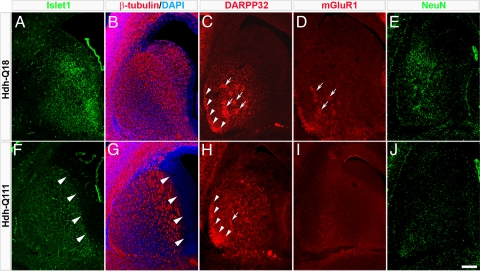
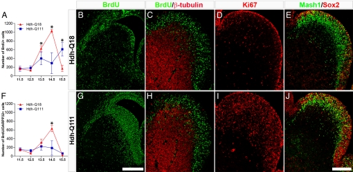
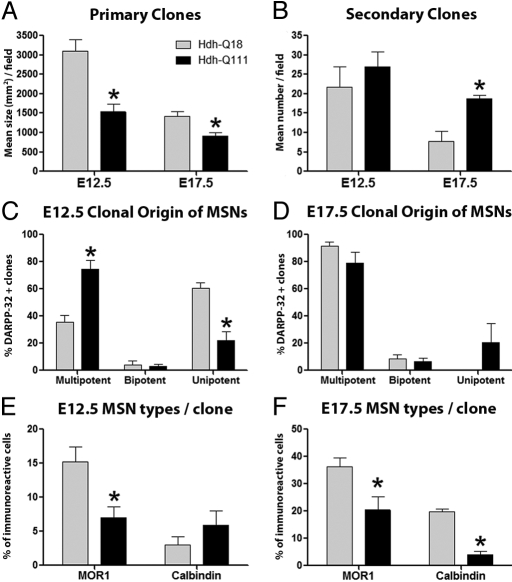
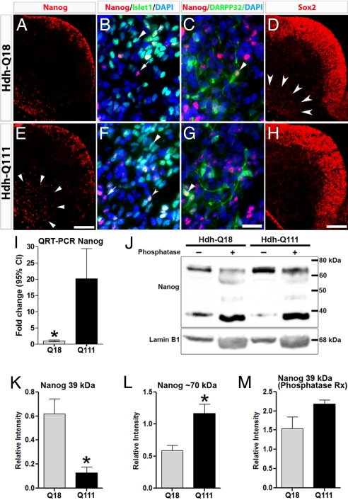
The pathogenesis of Huntington's disease (HD) remains elusive. The identification of increasingly early pathophysiological abnormalities in HD suggests the possibility that impairments of striatal medium spiny neuron (MSN) specification and maturation may underlie the etiology of HD. In fact, we demonstrate that HD knock-in (Hdh-Q111) mice exhibited delayed acquisition of early striatal cytoarchitecture with aberrant expression of progressive markers of MSN neurogenesis (Islet1, DARPP-32, mGluR1, and NeuN). Hdh-Q111 striatal progenitors also displayed delayed cell cycle exit between E13.5-15.5 (BrdU birth-dating) and an enhanced fraction of abnormal cycling cells in association with expansion of the pool of intermediate progenitors and over expression of the core pluripotency (PP) factor, Sox2. Clonal analysis further revealed that Hdh-Q111 neural stem cells (NSCs) displayed: impaired lineage restriction, reduced proliferative potential, enhanced late-stage self-renewal, and deregulated MSN subtype specification. Further, our analysis revealed that in addition to Sox2, the core PP factor, Nanog is expressed within the striatal generative and mantle regions, and in Hdh-Q111 embryos the fraction of Nanog-expressing MSN precursors was substantially increased. Moreover, compared to Hdh-Q18 embryos, the Hdh-Q111 striatal anlagen exhibited significantly higher levels of the essential PP cofactor, Stat3. These findings suggest that Sox2 and Nanog may play roles during a selective window of embryonic brain maturation, and alterations of these factors may, in part, be responsible for mediating the aberrant program of Hdh-Q111 striatal MSN specification and maturation. We propose that these HD-associated developmental abnormalities might compromise neuronal homeostasis and subsequently render MSNs more vulnerable to late life stressors.


Conflict of interest statement

The authors declare no conflict of interest.

Figures

Fig. 1.
Fig. 1.
Comparative immunofluorescence micrographs of the E17.5 striatum revealed impaired acquisition of the Hdh-Q111 striatal cytoarchitecture. Compared to Hdh-Q18 embryos, the expression of Islet1 (A and F) and β-tubulin (C and H) were reduced in the Hdh-Q111 striatal germinative region (arrowheads). DARPP32 expression in the Hdh-Q111 striatum was also poorly organized in patch clusters (C and H, arrows) and the subcallosal streak appeared to be immature (arrowheads). Hdh-Q111 embryos failed to exhibit the patchy distribution of mGluR1 (D and I, arrowheads) and NeuN expression was visibly reduced (E and J). (Scale bar, 200 μm.)
Fig. 2.
Fig. 2.
Hdh-Q111 embryos display abnormal profiles of striatal neurogenesis and cell cycle progression. BrdU birth-dating analysis was performed using BrdU pulse administration at different developmental intervals (from E11.5 through E15.5), and brains were then harvested at E17.5 (A and F). Striatal BrdU+ cells (A) and BrdU+/DARPP32+ (F) cells were quantified and the mean ± SEM. of at least three independent biological replicas represented. *, all P values correspond to <0.05. The number of Hdh-Q111 cells born between E13.5 and E14.5 was significantly reduced, whereas the number of Hdh-Q111 neurons born at E15.5 was significantly increased when compared with Hdh-Q18 profiles. (B and G) Illustrate representative samples of E15.5 BrdU birth-dating. The presence of E14.5 striatal cells in S-phase were assessed by BrdU pulse labeling followed by embryo harvesting 30 min later (C and H). Equivalent profiles of BrdU+ cells, both negative and positive for β-tubulin were observed between Hdh-Q18 and Hdh-Q111 embryos. Profiles of expression of striatal Ki67 (D and I) and Sox2/Mash1 (E and J) were assessed by immunostaining at E14.5. Both Ki67 and Sox2 expression were enhanced in Hdh-Q111 embryos. (Scale bar, 500 μm in G and 200 μm in J.)
Fig. 3.
Fig. 3.
Hdh-Q111 neural stem and progenitor cell species exhibit reduced proliferation potential, enhanced late-stage self-renewal and impaired generation of MSN subtypes. Clonal expansion assays revealed reduced mean size of E12.5 and E17.5 Hdh-Q111 primary clones (A), while stem cell self-renewal was enhanced in Hdh-Q111 secondary clones at E17.5 (B). The lineage composition of clones that give rise to MSNs (DARPP32+ cells) (C and D) was examined by quantifying the proportion of clones that expressed three (multipotent), two (bipotent) or one (unipotent) neural lineage markers: β-tubulin, O4, and GFAP. Hdh-Q111 MSNs originated predominantly from multipotent clones at E12.5 (C), whereas a substantial fraction of these cells originated from unipotent clones at E17.5 (D). Compared to the Hdh-Q18 model, the fractions of MOR1+ cells generated from early clones (E) and of both MOR1+ and CB+ cells generated from late clones (F) were significantly reduced in the Hdh-Q111 model. Bars in A–F represent the mean ± SEM. of at least four independent biological replicas; *, all P values correspond to <0.05.
Fig. 4.
Fig. 4.
Expression profiles of Nanog and Sox2 are deregulated in Hdh-Q111 embryos at E12.5. Nanog displayed comparable expression profiles in LGE germinative zones of both models (A and E), whereas in the Hdh-Q111 mantle Nanog expression was substantially increased (arrowheads). Within the mantle region, Nanog colocalizes with the MSN markers, Islet1 (B and F, arrowheads) and DARPP32 (C and G, arrowheads). Sox2 immunoreactivity in the E12.5 LGE generative zone was equivalent in both models (D and H), whereas Sox2 expression (arrowheads) was reduced in the Hdh-Q111 mantle. (Scale bar, 100 μm (E and H) and 20 μm (G).) QRT-PCR analysis revealed that Nanog transcript expression was significantly increased in Hdh-Q111 relative to Hdh-Q18 specimens at E12.5 (I). Bars in I represent the mean ± 95%CI of four independent biological replicas. Immunoblot analysis of Nanog in E12.5 specimens (J) demonstrated a lower relative intensity of the approximately 39-kDa band (K) and a higher relative intensity of the approximately 70-kDa band (L) in the Hdh-Q111 compared to Hdh-Q18 specimens, with normalization of the levels of expression of the 39-kDa band following phosphatase treatment (M). Bars in K–M represent the mean ± SEM. of four independent biological replicas. *, all P values correspond to <0.05.

References

    1. U.S.-Venezuela Collaborative Research Project. A novel gene containing a trinucleotide repeat that is expanded and unstable on Huntington's disease chromosomes. The Huntington's disease collaborative research group. Cell. 1993;72:971–983. - PubMed
    1. Strong TV, et al. Widespread expression of the human and rat Huntington's disease gene in brain and nonneural tissues. Nat Genet. 1993;5:259–265. - PubMed
    1. Vonsattel JP, et al. Neuropathological classification of Huntington's disease. J Neuropathol Exp Neurol. 1985;44:559–577. - PubMed
    1. Woda JM, et al. Inactivation of the Huntington's disease gene (Hdh) impairs anterior streak formation and early patterning of the mouse embryo. BMC Develop Biol. 2005;5:17. - PMC - PubMed
    1. Reiner A, et al. Neurons lacking huntingtin differentially colonize brain and survive in chimeric mice. J Neurosci. 2001;21:7608–7619. - PMC - PubMed

Publication types