Insulin-stimulated interaction with 14-3-3 promotes cytoplasmic localization of lipin-1 in adipocytes
- PMID: 19955570
- PMCID: PMC2823528
- DOI: 10.1074/jbc.M109.072488
Insulin-stimulated interaction with 14-3-3 promotes cytoplasmic localization of lipin-1 in adipocytes
Abstract
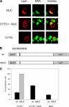
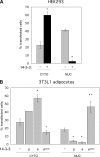
Lipin-1 is a bifunctional protein involved in lipid metabolism and adipogenesis. Lipin-1 plays a role in the biosynthesis of triacylglycerol through its phosphatidate phosphatase activity and also acts as a transcriptional co-activator of genes involved in oxidative metabolism. Lipin-1 resides in the cytoplasm and translocates to the endoplasmic reticulum membrane to catalyze the phosphatidate phosphatase reaction. It also possesses a nuclear localization signal, which is required for its translocation to the nucleus and may therefore be important for lipin-1 co-activator function. Thus, subcellular localization may be an important factor in the regulation of this protein. Here, we show that the nuclear localization signal alone is not sufficient for lipin-1 nuclear localization, and identify lipin-1 interaction with 14-3-3 as a determinant of its subcellular localization. We demonstrate that lipin-1 interacts with 14-3-3 proteins and that overexpression of 14-3-3 promotes the cytoplasmic localization of lipin-1 in 3T3-L1 adipocytes. The effect of 14-3-3 is mediated through a serine-rich domain in lipin-1. Functional mapping of the 14-3-3-interacting region within the serine-rich domain indicates redundancy and cooperativity among several sites, including five phosphorylated serine and threonine residues. Insulin stimulation of 3T3-L1 adipocytes results in increased lipin-1 phosphorylation, enhanced interaction with 14-3-3, and predominantly cytoplasmic localization. In summary, our studies suggest that insulin may modulate the cellular function of lipin-1 by regulating its subcellular localization through interactions with 14-3-3 proteins.
Figures


Similar articles
-
Conserved residues in the N terminus of lipin-1 are required for binding to protein phosphatase-1c, nuclear translocation, and phosphatidate phosphatase activity.J Biol Chem. 2014 Apr 11;289(15):10876-10886. doi: 10.1074/jbc.M114.552612. Epub 2014 Feb 20. J Biol Chem. 2014. PMID: 24558042 Free PMC article.
-
Casein kinase II-mediated phosphorylation of lipin 1β phosphatidate phosphatase at Ser-285 and Ser-287 regulates its interaction with 14-3-3β protein.J Biol Chem. 2019 Feb 15;294(7):2365-2374. doi: 10.1074/jbc.RA118.007246. Epub 2019 Jan 7. J Biol Chem. 2019. PMID: 30617183 Free PMC article.
-
PPARγ agonists induce adipocyte differentiation by modulating the expression of Lipin-1, which acts as a PPARγ phosphatase.Int J Biochem Cell Biol. 2016 Dec;81(Pt A):57-66. doi: 10.1016/j.biocel.2016.10.018. Epub 2016 Oct 22. Int J Biochem Cell Biol. 2016. PMID: 27780754
-
Lipin - The bridge between hepatic glycerolipid biosynthesis and lipoprotein metabolism.Biochim Biophys Acta. 2010 Dec;1801(12):1249-59. doi: 10.1016/j.bbalip.2010.07.008. Epub 2010 Aug 6. Biochim Biophys Acta. 2010. PMID: 20692363 Review.
-
The lipin protein family: dual roles in lipid biosynthesis and gene expression.FEBS Lett. 2008 Jan 9;582(1):90-6. doi: 10.1016/j.febslet.2007.11.014. Epub 2007 Nov 20. FEBS Lett. 2008. PMID: 18023282 Free PMC article. Review.
Cited by
-
Conserved residues in the N terminus of lipin-1 are required for binding to protein phosphatase-1c, nuclear translocation, and phosphatidate phosphatase activity.J Biol Chem. 2014 Apr 11;289(15):10876-10886. doi: 10.1074/jbc.M114.552612. Epub 2014 Feb 20. J Biol Chem. 2014. PMID: 24558042 Free PMC article.
-
The middle lipin domain adopts a membrane-binding dimeric protein fold.Nat Commun. 2021 Aug 5;12(1):4718. doi: 10.1038/s41467-021-24929-5. Nat Commun. 2021. PMID: 34354069 Free PMC article.
-
The phosphatidic acid-binding, polybasic domain is responsible for the differences in the phosphoregulation of lipins 1 and 3.J Biol Chem. 2017 Dec 15;292(50):20481-20493. doi: 10.1074/jbc.M117.786574. Epub 2017 Oct 5. J Biol Chem. 2017. PMID: 28982975 Free PMC article.
-
Metabolic Alterations in Myotonic Dystrophy Type 1 and Their Correlation with Lipin.Int J Environ Res Public Health. 2021 Feb 12;18(4):1794. doi: 10.3390/ijerph18041794. Int J Environ Res Public Health. 2021. PMID: 33673200 Free PMC article. Review.
-
The Role of Torsin AAA+ Proteins in Preserving Nuclear Envelope Integrity and Safeguarding Against Disease.Biomolecules. 2020 Mar 19;10(3):468. doi: 10.3390/biom10030468. Biomolecules. 2020. PMID: 32204310 Free PMC article. Review.
References
Publication types
MeSH terms
Substances
Grants and funding
LinkOut - more resources
Full Text Sources
Other Literature Sources
Medical
Molecular Biology Databases

