. 2010 Jan;120(1):290-302.
doi: 10.1172/JCI39194. Epub 2009 Dec 1.

TGF-beta1-induced expression of human Mdm2 correlates with late-stage metastatic breast cancer

Affiliations

TGF-beta1-induced expression of human Mdm2 correlates with late-stage metastatic breast cancer

Shinako Araki et al. J Clin Invest. 2010 Jan.

Abstract

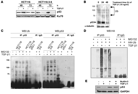
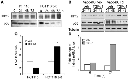
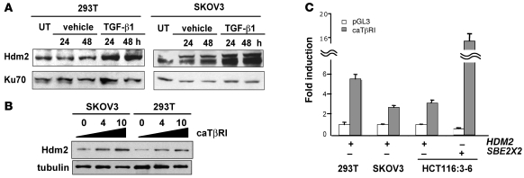
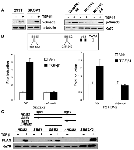
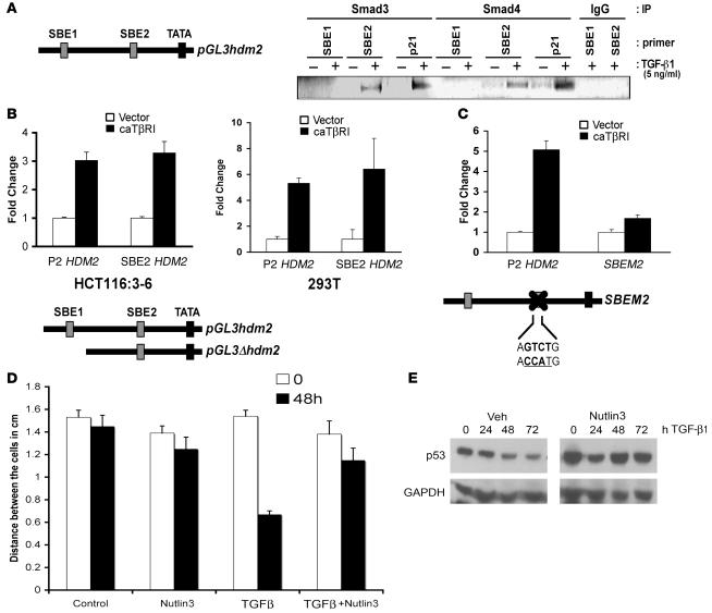
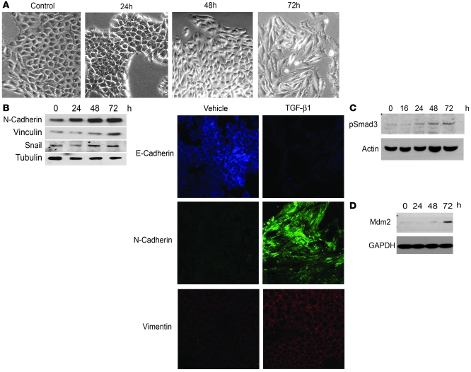
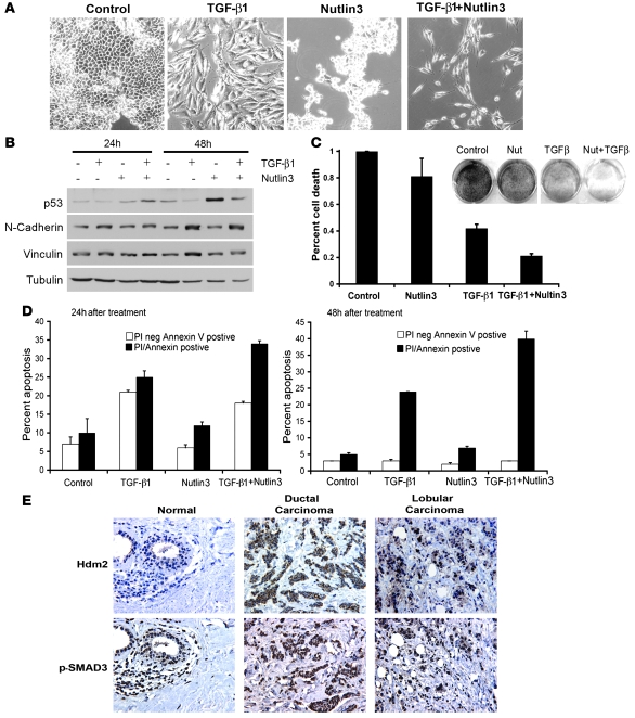
The E3 ubiquitin ligase human murine double minute (HDM2) is overexpressed in 40%-80% of late-stage metastatic cancers in the absence of gene amplification. Hdm2 regulates p53 stability via ubiquitination and has also been implicated in altering the sensitivity of cells to TGF-beta1. Whether TGF-beta1 signaling induces Hdm2 expression leading to HDM2-mediated destabilization of p53 has not been investigated. In this study, we report that TGF-beta1-activated SMA- and MAD3 (Smad3/4) transcription factors specifically bound to the second promoter region of HDM2, leading to increased HDM2 protein expression and destabilization of p53 in human cancer cell lines. Additionally, TGF-beta1 expression led to Smad3 activation and murine double minute 2 (Mdm2) expression in murine mammary epithelial cells during epithelial-to-mesenchymal transition (EMT). Furthermore, histological analyses of human breast cancer samples demonstrated that approximately 65% of late-stage carcinomas were positive for activated Smad3 and HDM2, indicating a strong correlation between TGF-beta1-mediated induction of HDM2 and late-stage tumor progression. Identification of Hdm2 as a downstream target of TGF-beta1 represents a critical prosurvival mechanism in cancer progression and provides another point for therapeutic intervention in late-stage cancer.


Figures

Figure 1
Figure 1. p53 destabilization by TGF-β1 stimulation.
(A) HCT116 and HCT116:3-6 cells were treated with vehicle or 10 ng/ml of TGF-β1 for 48 and 72 hours. Cellular extracts were prepared for Western blot, and p53 and Ku70 (internal control) levels were detected. (B) Western blot of p53 laddering, an indication of ubiquitination (Ub) (arrows) in cellular extracts of HCT116:3-6 cells treated with 10 ng/ml of TGF-β1 for 0, 24, and 48 hours. (C) HCT116, Hct116 p53–/–, and HCT116:3-6 cells were treated with 10 ng/ml of TGF-β1 with or without the proteasome inhibitor MG132 (30 μM). Cellular extracts were immunoprecipitated with control IgG or p53 antibodies. Precipitates were separated on an SDS-PAGE gel, and a Western blot was prepared. The left side was probed for ubiquitin. The right side was probed for p53. (D) HCT116:3-6 cells were transfected with HA-Ub. Cells were then treated with MG132 (30 μM) with or without TGF-β1 (10 ng/ml); cellular extracts were immunoprecipitated with p53 or IgG antibodies and prepared for Western blot analysis of HA. (E) Western blot of p53 in extracts isolated from HCT116:3-6 cells treated with TGF-β1 (10 ng/ml) with or without Nutlin3 (10 μM).
Figure 2
Figure 2. TGF-β1 increases Hdm2 mRNA and protein levels.
(A) Western blot analysis of Hdm2 and p53 in HCT116 and HCT116:3-6 treated with TGF-β1 (10 ng/ml) for 0, 24, 48, and 72 hours. (B) Vaco400 cells with control plasmid (Vaco400:neo) or expressing the functional TGF-β1 receptor II (Vaco400:RII) cells were treated with vehicle (veh) or TGF-β1 (10 ng/ml) for 24 or 48 hours. Western blot was prepared from the extracts, and Hdm2, p53, and tubulin were detected. (C) HCT116 and HCT116:3-6 were transfected with the HDM2 promoter-luciferase reporter construct and renilla expression plasmid; then cells were treated with TGF-β1 (10 ng/ml) or vehicle. After 48 hours, extracts were prepared for analysis. Fold induction represents vehicle to TGF-β1 treatments and error bars represent SD generated from the mean. (D) Real-time PCR was performed on HCT116:3-6 cells treated with vehicle or TGF-β1 (10 ng/ml) at 6 or 24 hours.
Figure 3
Figure 3. Increased Hdm2 in response to TGF-β1 is independent of p53.
(A) 293T and SKOV3 cells were untreated (UT) or treated with vehicle or TGF-β1 (10 ng/ml) for 24 and 48 hours. Cellular extracts were prepared for Western blot analysis of Hdm2 and Ku70. (B) SKOV3 and 293T cells were transiently transfected with 0, 4, or 10 μg of constitutively active TβRI (caTβRI) expression plasmid. Cellular extracts were prepared for Western blot analysis of Hdm2 and α-tubulin. (C) Transient transfection of 293T, SKOV3, and HCT116:3-6 cells with the caTβRI and HDM2 reporter or the SBE2X2 reporter construct. All samples were transfected with a renilla construct. Reporter activity was determined relative to renilla to generate relative activity. Fold induction was determined relative to vehicle control and SD was calculated relative to the mean.
Figure 4
Figure 4. Smad activation and induction of the HDM2 promoter.
(A) Western blot of p-Smad3 in 293T, SKOV3, Vaco400:RII, HCT116, and HCT116:3-6 cells. Tubulin and Ku70 were used as internal controls for loading. (B) A schematic of 2 putative SBEs designated SBE1 and SBE2 and the p53-binding elements in the P2 promoter of HDM2. Reporter assays were conducted using dominant negative Smad4 (dnSmad4) or control vector only (VO) in transient transfection assays. HCT116:3-6 cells were transfected with an SBE2X2 reporter as a positive control or HDM2 P2 reporter. All samples were transfected with renilla to use as an internal control. Cells were treated with vehicle or TGF-β1 (10 ng/ml). Reporter activity was determined relative to renilla to generate relative activity. Fold induction was determined relative to vehicle control and SD was calculated relative to the mean. (C) Western blot of FLAG-Smad3 and Ku70 that bound to the HDM2 promoter. 293T cells were transfected with FLAG-Smad3 and treated with vehicle or TGF-β1 (10 ng/ml). Nuclear extracts were prepared from the latter mentioned cell treatments. Biotinylated PCR fragments of the HDM2 promoter, SBE1, SBE2, ΔHDM2, or a positive control, SBE2X2, were mixed with the nuclear extracts. Biotinylated DNA fragments bound to streptavidin beads were used to purify FLAG-Smad3 and Ku70 (internal control).
Figure 5
Figure 5. Binding of Smad3 and Smad4 to the HDM2 SBE2 in vivo.
(A) HCT116:3-6 cells were treated with vehicle or TGF-β1 (10 ng/ml) for 48 hours. Smad3, Smad4, or IgG isotype control antibodies were used to immunoprecipitate Smads binding to the HDM2 promoter. Oligo sets were used to PCR amplify the SBE1, SBE2, or the p21 promoter SBE (a positive control for Smad3/4 binding) and resolved on an agarose gel. (B) HCT116:3-6 and 293T cells were transfected with caTβRI and either the full-length P2 HDM2-promoter driving luciferase or a deletion construct missing SBE1. Reporter activity was determined relative to renilla to generate relative activity. Fold induction was determined relative to vehicle control, and SD was calculated relative to the mean. (C) Cells were transfected with caTβRI and the P2 HDM2 promoter driving luciferase or a mutant whereby the SBE2 site was mutated. A renilla expression construct was used as an internal control for normalization. SEM was calculated as in B. (D) Wound healing assay using HCT116:3-6 cells, scored in a double-blind assay. Bar graph represents the distance between confluent cells after stimulation with TGF-β1 (10 ng/ml), Nutlin3 (10 μM), or TGF-β1 (10 ng/ml) and Nutlin3 (10 μM). (E) Western blot analysis of p53 in HCT116:3-6 cells treated with TGF-β1 (10 ng/ml) with or without Nutlin3 (10 μM) over a 72-hour time course. SD was calculated relative to the mean.
Figure 6
Figure 6. Characterization of TGF-β1 treatment on cell migration and EMT.
(A) NMuMg mammary cells treated with 4 ng/ml of TGF-β1. Photographs were taken at 0, 24, 48, and 72 hours. (B) N-cadherin, Vinculin, Snail, and α-tubulin (loading control) were detected by Western blot of whole-cell lysates from NMuMg cells treated with TGF-β1 (4 ng/ml) over a 72-hour time course. Confocal microscopy of E-cadherin, N-cadherin, and Vimentin in NMuMG cells treated with 4 ng/ml. Original magnification, ×20. (C) Western blot of a time-course activation of p-Smad3 in NMuMg in response to TGF-β1 (4 ng/ml). Loading was normalized to actin. (D) Time-course analysis of Mdm2 induction as measured by Western blot after treatment of NMuMg cells with TGF-β1 (4 ng/ml).
Figure 7
Figure 7. Induction of apoptosis in response to TGF-β1 and Nutlin3 exposure.
(A) Morphological changes to NMuMg cells after incubation with 4 ng/ml TGF-β1, Nutlin3 (10 μM), or TGF-β1 and Nutlin3. Original magnification, ×20. (B) Western blot of p53, N-cadherin, Vinculin, and Tubulin under the same treatment conditions as A. (C) Survival assay of NMuMg cells. Cell were treated as in A and stained with methylene blue at day 5. Methylene blue was liberated from the cells and quantified by OD595. Percentage of cell death was calculated from control; data represent 3 independent experiments completed in triplicate. SD was calculated relative to the mean. (D) Flow cytometry of NMuMg cells treated as in A and stained for annexin V and PI at 24 and 48 hours. Data represent triplicates of experiments. White bars represent PI negative and annexin V positive, and black represents PI and annexin V positive. SD was calculated relative to the mean. (E) Immunohistochemistry of Hdm2 and p-Smad3 (serine 425) in normal breast tissue, ductal carcinoma, and lobular carcinoma. Original magnification, ×16.

Similar articles

Cited by

References

    1. Roberts AB, Wakefield LM. The two faces of transforming growth factor beta in carcinogenesis. Proc Natl Acad Sci U S A. 2003;100(15):8621–8623. doi: 10.1073/pnas.1633291100. - DOI - PMC - PubMed
    1. Lu SL, et al. Overexpression of transforming growth factor beta1 in head and neck epithelia results in inflammation, angiogenesis, and epithelial hyperproliferation. Cancer Res. 2004;64(13):4405–4410. doi: 10.1158/0008-5472.CAN-04-1032. - DOI - PubMed
    1. Gold LI. The role for transforming growth factor-beta (TGF-beta) in human cancer. Crit Rev Oncog. 1999;10(4):303–360. - PubMed
    1. Derynck R, Zhang YE. Smad-dependent and Smad-independent pathways in TGF-beta family signalling. Nature. 2003;425(6958):577–584. doi: 10.1038/nature02006. - DOI - PubMed
    1. Heldin CH, Miyazono K, ten Dijke P. TGF-beta signalling from cell membrane to nucleus through SMAD proteins. Nature. 1997;390(6659):465–471. doi: 10.1038/37284. - DOI - PubMed

Publication types

MeSH terms